[English] 日本語
Yorodumi
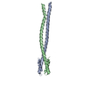
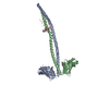
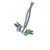
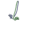
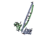
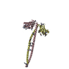
- PDB-5cj4: Crystal Structure of Amino Acids 1562-1622 of MYH7 -

+
Open data


ID or keywords:

Loading...

-
Basic information

Entry
Database: PDB / ID: 5cj4
TitleCrystal Structure of Amino Acids 1562-1622 of MYH7
ComponentsXrcc4-MYH7-(1562-1622) chimera protein
KeywordsMOTOR PROTEIN / Myosin / coiled-coil
Function / homology
Function and homology information


FHA domain binding / regulation of slow-twitch skeletal muscle fiber contraction / regulation of the force of skeletal muscle contraction / positive regulation of ligase activity / DNA ligase IV complex / DNA ligation involved in DNA repair / muscle myosin complex / muscle filament sliding / DNA-dependent protein kinase-DNA ligase 4 complex / immunoglobulin V(D)J recombination ...FHA domain binding / regulation of slow-twitch skeletal muscle fiber contraction / regulation of the force of skeletal muscle contraction / positive regulation of ligase activity / DNA ligase IV complex / DNA ligation involved in DNA repair / muscle myosin complex / muscle filament sliding / DNA-dependent protein kinase-DNA ligase 4 complex / immunoglobulin V(D)J recombination / nonhomologous end joining complex / regulation of the force of heart contraction / protein localization to site of double-strand break / transition between fast and slow fiber / myosin filament / myosin II complex / adult heart development / cardiac muscle hypertrophy in response to stress / myosin complex / sarcomere organization / microfilament motor activity / ventricular cardiac muscle tissue morphogenesis / myofibril / cellular response to lithium ion / 2-LTR circle formation / skeletal muscle contraction / response to X-ray / striated muscle contraction / SUMOylation of DNA damage response and repair proteins / stress fiber / ATP metabolic process / cardiac muscle contraction / regulation of heart rate / sarcomere / muscle contraction / Nonhomologous End-Joining (NHEJ) / Z disc / double-strand break repair via nonhomologous end joining / double-strand break repair / actin filament binding / site of double-strand break / calmodulin binding / nucleoplasm / ATP binding / identical protein binding / nucleus / cytosol / cytoplasm
Similarity search - Function
Single alpha-helices involved in coiled-coils or other helix-helix interfaces - #370 / DNA double-strand break repair and VJ recombination XRCC4, N-terminal / Dna Repair Protein Xrcc4; Chain: A, domain 1 / XRCC4, N-terminal domain superfamily / DNA repair protein XRCC4 / DNA double-strand break repair and V(D)J recombination protein XRCC4 / XRCC4-like, N-terminal domain superfamily / DNA repair protein XRCC4-like, C-terminal / Myosin tail / Myosin tail ...Single alpha-helices involved in coiled-coils or other helix-helix interfaces - #370 / DNA double-strand break repair and VJ recombination XRCC4, N-terminal / Dna Repair Protein Xrcc4; Chain: A, domain 1 / XRCC4, N-terminal domain superfamily / DNA repair protein XRCC4 / DNA double-strand break repair and V(D)J recombination protein XRCC4 / XRCC4-like, N-terminal domain superfamily / DNA repair protein XRCC4-like, C-terminal / Myosin tail / Myosin tail / Myosin N-terminal SH3-like domain / Myosin S1 fragment, N-terminal / Myosin, N-terminal, SH3-like / Myosin N-terminal SH3-like domain profile. / Short calmodulin-binding motif containing conserved Ile and Gln residues. / Myosin head, motor domain / Myosin head (motor domain) / Myosin motor domain profile. / Myosin. Large ATPases. / IQ motif profile. / IQ motif, EF-hand binding site / Kinesin motor domain superfamily / Beta Complex / Single alpha-helices involved in coiled-coils or other helix-helix interfaces / Up-down Bundle / P-loop containing nucleoside triphosphate hydrolase / Mainly Beta / Mainly Alpha
Similarity search - Domain/homology
Myosin-7 / DNA repair protein XRCC4
Similarity search - Component
Biological speciesHomo sapiens (human)
MethodX-RAY DIFFRACTION / SYNCHROTRON / MOLECULAR REPLACEMENT / Resolution: 3.102 Å
AuthorsKorkmaz, N.E. / Taylor, K.C. / Andreas, M.P. / Ajay, G. / Heinze, N.T. / Cui, Q. / Rayment, I.
Funding support United States, 2items
OrganizationGrant numberCountry
National Institutes of Health/National Heart, Lung, and Blood Institute (NIH/NHLBI)HL111237 United States
National Science Foundation (NSF, United States)CHE1300209 United States
CitationJournal: Proteins / Year: 2016
Title: A composite approach towards a complete model of the myosin rod.
Authors: Korkmaz, E.N. / Taylor, K.C. / Andreas, M.P. / Ajay, G. / Heinze, N.T. / Cui, Q. / Rayment, I.
History
DepositionJul 13, 2015Deposition site: RCSB / Processing site: RCSB
Revision 1.0Dec 2, 2015Provider: repository / Type: Initial release
Revision 1.1Jan 27, 2016Group: Database references
Revision 1.2Sep 27, 2017Group: Author supporting evidence / Database references / Derived calculations
Category: citation / pdbx_audit_support / pdbx_struct_oper_list
Item: _citation.journal_id_CSD / _pdbx_audit_support.funding_organization / _pdbx_struct_oper_list.symmetry_operation
Revision 1.3Nov 27, 2019Group: Author supporting evidence / Category: pdbx_audit_support / Item: _pdbx_audit_support.funding_organization
Revision 1.4Sep 27, 2023Group: Data collection / Database references / Refinement description
Category: chem_comp_atom / chem_comp_bond ...chem_comp_atom / chem_comp_bond / database_2 / pdbx_initial_refinement_model / struct_ncs_dom_lim
Item: _database_2.pdbx_DOI / _database_2.pdbx_database_accession ..._database_2.pdbx_DOI / _database_2.pdbx_database_accession / _struct_ncs_dom_lim.beg_auth_comp_id / _struct_ncs_dom_lim.beg_label_asym_id / _struct_ncs_dom_lim.beg_label_comp_id / _struct_ncs_dom_lim.beg_label_seq_id / _struct_ncs_dom_lim.end_auth_comp_id / _struct_ncs_dom_lim.end_label_asym_id / _struct_ncs_dom_lim.end_label_comp_id / _struct_ncs_dom_lim.end_label_seq_id

-
Structure visualization

Structure viewerMolecule:
MolmilJmol/JSmol

Downloads & links

-
Assembly

Deposited unit
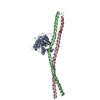
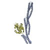
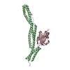
A: Xrcc4-MYH7-(1562-1622) chimera protein
B: Xrcc4-MYH7-(1562-1622) chimera protein
C: Xrcc4-MYH7-(1562-1622) chimera protein
D: Xrcc4-MYH7-(1562-1622) chimera protein


Theoretical massNumber of molelcules
Total (without water)96,4294
Polymers96,4294
Non-polymers00
Water362
1
A: Xrcc4-MYH7-(1562-1622) chimera protein
B: Xrcc4-MYH7-(1562-1622) chimera protein


Theoretical massNumber of molelcules
Total (without water)48,2152
Polymers48,2152
Non-polymers00
Water181
TypeNameSymmetry operationNumber
identity operation1_555x,y,z1
Buried area4700 Å2
ΔGint-44 kcal/mol
Surface area23260 Å2
MethodPISA
2
C: Xrcc4-MYH7-(1562-1622) chimera protein
D: Xrcc4-MYH7-(1562-1622) chimera protein


Theoretical massNumber of molelcules
Total (without water)48,2152
Polymers48,2152
Non-polymers00
Water181
TypeNameSymmetry operationNumber
identity operation1_555x,y,z1
Buried area5030 Å2
ΔGint-46 kcal/mol
Surface area23660 Å2
MethodPISA
Unit cell
Length a, b, c (Å)83.145, 57.285, 112.100
Angle α, β, γ (deg.)90.000, 99.860, 90.000
Int Tables number4
Space group name H-MP1211
Noncrystallographic symmetry (NCS)NCS domain:
IDEns-IDDetails
11chain A
21chain B
31chain C
41chain D

NCS domain segments:

Component-ID: 1 / Ens-ID: 1 / Beg auth comp-ID: GLY / Beg label comp-ID: GLY

Dom-IDEnd auth comp-IDEnd label comp-IDSelection detailsAuth asym-IDLabel asym-IDAuth seq-IDLabel seq-ID
1MLYMLYchain AAA1 - 16154 - 201
2ARGARGchain BBB1 - 16084 - 194
3GLUGLUchain CCC1 - 16194 - 205
4LEULEUchain DDD1 - 16124 - 198

-
Components

#1: Protein
Xrcc4-MYH7-(1562-1622) chimera protein / X-ray repair cross-complementing protein 4 / Myosin heavy chain 7 / Myosin heavy chain slow isoform ...X-ray repair cross-complementing protein 4 / Myosin heavy chain 7 / Myosin heavy chain slow isoform / MyHC-slow / Myosin heavy chain / cardiac muscle beta isoform / MyHC-beta


Mass: 24107.324 Da / Num. of mol.: 4 / Fragment: UNP Q13426 residues 2-144, UNP P12883 1562-1622
Source method: isolated from a genetically manipulated source
Details: Lysines were reductively alkylated to dimethyllysine.
Source: (gene. exp.) Homo sapiens (human) / Gene: XRCC4, MYH7, MYHCB / Production host: Escherichia coli (E. coli) / Strain (production host): BL21(DE3) / References: UniProt: Q13426, UniProt: P12883
#2: Water ChemComp-HOH / water / Water


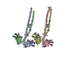
Mass: 18.015 Da / Num. of mol.: 2 / Source method: isolated from a natural source / Formula: H2O

-
Experimental details

-
Experiment

ExperimentMethod: X-RAY DIFFRACTION / Number of used crystals: 1

-
Sample preparation

CrystalDensity Matthews: 2.73 Å3/Da / Density % sol: 54.91 %
Crystal growTemperature: 277 K / Method: vapor diffusion, hanging drop / pH: 8
Details: 23% (w/v) PEG 4000, 500 mM NaCl, 100 mM triethanolamine pH 8.0.

-
Data collection

DiffractionMean temperature: 100 K
Diffraction sourceSource: SYNCHROTRON / Site: APS / Beamline: 19-ID / Wavelength: 0.97918 Å
DetectorType: ADSC QUANTUM 315r / Detector: CCD / Date: Aug 9, 2014
RadiationMonochromator: Rosenbaum-Rock high-resolution double-crystal monochromator. LN2 cooled first crystal, sagittal focusing 2nd crystal
Protocol: SINGLE WAVELENGTH / Monochromatic (M) / Laue (L): M / Scattering type: x-ray
Radiation wavelengthWavelength: 0.97918 Å / Relative weight: 1
ReflectionResolution: 3.1→50 Å / Num. obs: 19051 / % possible obs: 99.1 % / Redundancy: 6 % / Biso Wilson estimate: 66.66 Å2 / Rmerge(I) obs: 0.07 / Net I/σ(I): 27.2
Reflection shellResolution: 3.1→3.15 Å / Redundancy: 5.9 % / Rmerge(I) obs: 0.366 / Mean I/σ(I) obs: 4.9 / % possible all: 99.4

-
Processing

Software
NameVersionClassification
PHENIX1.9_1692refinement
PDB_EXTRACT3.15data extraction
HKL-3000data reduction
HKL-3000data scaling
PHENIX1.9_1692phasing
RefinementMethod to determine structure: MOLECULAR REPLACEMENT
Starting model: 1IK9
Resolution: 3.102→49.923 Å / SU ML: 0.41 / Cross valid method: FREE R-VALUE / σ(F): 1.36 / Phase error: 31.14 / Stereochemistry target values: ML
RfactorNum. reflection% reflection
Rfree0.2796 948 5 %
Rwork0.2432 18011 -
obs0.245 18959 98.6 %
Solvent computationShrinkage radii: 0.9 Å / VDW probe radii: 1.11 Å / Solvent model: FLAT BULK SOLVENT MODEL
Displacement parametersBiso max: 191.26 Å2 / Biso mean: 81.9698 Å2 / Biso min: 35.56 Å2
Refinement stepCycle: final / Resolution: 3.102→49.923 Å
ProteinNucleic acidLigandSolventTotal
Num. atoms6341 0 0 2 6343
Biso mean---61.58 -
Num. residues----770
Refine LS restraints
Refine-IDTypeDev idealNumber
X-RAY DIFFRACTIONf_bond_d0.0046433
X-RAY DIFFRACTIONf_angle_d0.7418655
X-RAY DIFFRACTIONf_chiral_restr0.032940
X-RAY DIFFRACTIONf_plane_restr0.0031098
X-RAY DIFFRACTIONf_dihedral_angle_d14.5862447
Refine LS restraints NCS
Ens-IDDom-IDAuth asym-IDNumberRefine-IDRmsType
11A3056X-RAY DIFFRACTION12.111TORSIONAL
12B3056X-RAY DIFFRACTION12.111TORSIONAL
13C3056X-RAY DIFFRACTION12.111TORSIONAL
14D3056X-RAY DIFFRACTION12.111TORSIONAL
LS refinement shell

Refine-ID: X-RAY DIFFRACTION / Total num. of bins used: 7

Resolution (Å)Rfactor RfreeNum. reflection RfreeRfactor RworkNum. reflection RworkNum. reflection all% reflection obs (%)
3.1016-3.26510.32411320.28442520265298
3.2651-3.46960.32361340.28542564269899
3.4696-3.73740.29231360.25332579271599
3.7374-4.11330.24931350.24932562269799
4.1133-4.70820.25711350.21622572270799
4.7082-5.93020.26941380.2392587272598
5.9302-49.92920.28841380.2332627276598
Refinement TLS params.

Method: refined / Refine-ID: X-RAY DIFFRACTION

IDL112)L122)L132)L222)L232)L332)S11 (Å °)S12 (Å °)S13 (Å °)S21 (Å °)S22 (Å °)S23 (Å °)S31 (Å °)S32 (Å °)S33 (Å °)T112)T122)T132)T222)T232)T332)Origin x (Å)Origin y (Å)Origin z (Å)
13.7725-0.3135-1.42931.80221.96345.21350.00290.3266-0.1993-0.2331-0.1334-0.1818-0.14590.9860.14840.45760.01-0.03190.72330.04670.535896.3493-17.7439110.69
26.16560.90322.5315-0.23730.14781.99950.21960.19031.49710.0922-0.75780.5431-0.22-0.50450.30040.47280.0961-0.19181.1474-0.17290.781469.4668-15.4385118.3763
37.16871.58991.87522.91551.13475.22380.687-0.7172-0.96250.327-0.4434-0.06251.1442-0.496-0.00550.6119-0.0064-0.07471.14180.00430.560519.8274-22.214989.8215
43.16640.2275-1.3872.2649-0.10074.37580.1073-0.4124-0.26650.305-0.36370.31820.4629-0.5090.10090.5017-0.17420.01890.6679-0.10140.668272.4945-22.9779135.0062
55.96120.22332.28012.87731.023.9635-0.0862-0.07680.9717-0.29320.13850.30220.3542-0.1289-0.25450.5127-0.0085-0.07090.9210.04320.563828.2548-16.307884.7998
63.4613-1.0392-2.15342.17940.94014.40880.05710.13750.281-0.32630.2343-0.0499-0.35690.1152-0.2140.5268-0.0785-0.05910.3972-0.01440.5013104.68339.377761.5806
71.01250.7805-0.82471.83491.07533.33390.0967-0.10860.5138-0.25490.1797-0.0516-0.01980.1667-0.13580.6369-0.0137-0.0290.40940.04910.45993.85528.555365.7969
82.92480.681-0.58052.25042.48491.6769-0.12830.15060.83910.0835-0.1980.2962-0.0650.30490.13560.7034-0.146-0.03330.83480.21840.506626.88920.465238.7101
94.4914-0.9013-1.8286-0.00280.28243.5608-0.2083-0.5317-0.04360.06390.23020.34150.1986-0.6775-0.05820.6087-0.0198-0.01440.8020.10020.698982.88395.448786.0213
108.95950.88652.40081.64660.49563.84060.5878-0.22930.27950.2863-0.34350.37160.90680.1699-0.04740.5269-0.0653-0.03440.76640.14690.591135.36194.730234.5294
Refinement TLS group
IDRefine-IDRefine TLS-IDSelection detailsAuth asym-IDAuth seq-ID
1X-RAY DIFFRACTION1chain 'A' and (resid 1 through 118 )A0
2X-RAY DIFFRACTION2chain 'A' and (resid 119 through 1572 )A0
3X-RAY DIFFRACTION3chain 'A' and (resid 1573 through 1614 )A0
4X-RAY DIFFRACTION4chain 'B' and (resid 1 through 1570 )B0
5X-RAY DIFFRACTION5chain 'B' and (resid 1571 through 1608 )B0
6X-RAY DIFFRACTION6chain 'C' and (resid 1 through 89 )C0
7X-RAY DIFFRACTION7chain 'C' and (resid 91 through 1572 )C0
8X-RAY DIFFRACTION8chain 'C' and (resid 1573 through 1619 )C0
9X-RAY DIFFRACTION9chain 'D' and (resid 1 through 1572 )D0
10X-RAY DIFFRACTION10chain 'D' and (resid 1573 through 1612 )D0

+
About Yorodumi

-
News

-
Feb 9, 2022. New format data for meta-information of EMDB entries

New format data for meta-information of EMDB entries

  • Version 3 of the EMDB header file is now the official format.
  • The previous official version 1.9 will be removed from the archive.

Related info.:EMDB header

External links:wwPDB to switch to version 3 of the EMDB data model

-
Aug 12, 2020. Covid-19 info

Covid-19 info

URL: https://pdbj.org/emnavi/covid19.php

New page: Covid-19 featured information page in EM Navigator.

Related info.:Covid-19 info / Mar 5, 2020. Novel coronavirus structure data

+
Mar 5, 2020. Novel coronavirus structure data

Novel coronavirus structure data

Related info.:Yorodumi Speices / Aug 12, 2020. Covid-19 info

External links:COVID-19 featured content - PDBj / Molecule of the Month (242):Coronavirus Proteases

+
Jan 31, 2019. EMDB accession codes are about to change! (news from PDBe EMDB page)

EMDB accession codes are about to change! (news from PDBe EMDB page)

  • The allocation of 4 digits for EMDB accession codes will soon come to an end. Whilst these codes will remain in use, new EMDB accession codes will include an additional digit and will expand incrementally as the available range of codes is exhausted. The current 4-digit format prefixed with “EMD-” (i.e. EMD-XXXX) will advance to a 5-digit format (i.e. EMD-XXXXX), and so on. It is currently estimated that the 4-digit codes will be depleted around Spring 2019, at which point the 5-digit format will come into force.
  • The EM Navigator/Yorodumi systems omit the EMD- prefix.

Related info.:Q: What is EMD? / ID/Accession-code notation in Yorodumi/EM Navigator

External links:EMDB Accession Codes are Changing Soon! / Contact to PDBj

+
Jul 12, 2017. Major update of PDB

Major update of PDB

  • wwPDB released updated PDB data conforming to the new PDBx/mmCIF dictionary.
  • This is a major update changing the version number from 4 to 5, and with Remediation, in which all the entries are updated.
  • In this update, many items about electron microscopy experimental information are reorganized (e.g. em_software).
  • Now, EM Navigator and Yorodumi are based on the updated data.

External links:wwPDB Remediation / Enriched Model Files Conforming to OneDep Data Standards Now Available in the PDB FTP Archive

-
Yorodumi

Thousand views of thousand structures

  • Yorodumi is a browser for structure data from EMDB, PDB, SASBDB, etc.
  • This page is also the successor to EM Navigator detail page, and also detail information page/front-end page for Omokage search.
  • The word "yorodu" (or yorozu) is an old Japanese word meaning "ten thousand". "mi" (miru) is to see.

Related info.:EMDB / PDB / SASBDB / Comparison of 3 databanks / Yorodumi Search / Aug 31, 2016. New EM Navigator & Yorodumi / Yorodumi Papers / Jmol/JSmol / Function and homology information / Changes in new EM Navigator and Yorodumi

Read more