 Movie
Movie Controller


+ Open data

- Basic information
| Entry | Database: PDB / ID: 4xa4 | ||||||
|---|---|---|---|---|---|---|---|
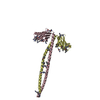
| Title | Crystal Structure of the coiled-coil surrounding Skip 3 of MYH7 | ||||||
 Components Components | Xrcc4-MYH7(1551-1609) chimera protein | ||||||
 Keywords Keywords | MOTOR PROTEIN / Myosin / coiled coil / skip residue / fusion / Gp7 / EB1 / MYH7 / Cardiac | ||||||
| Function / homology |  Function and homology information Function and homology informationFHA domain binding / regulation of slow-twitch skeletal muscle fiber contraction / regulation of the force of skeletal muscle contraction / positive regulation of ligase activity / DNA ligase IV complex / DNA double-strand break attachment to nuclear envelope / DNA-dependent protein kinase-DNA ligase 4 complex / muscle myosin complex / immunoglobulin V(D)J recombination / nonhomologous end joining complex ...FHA domain binding / regulation of slow-twitch skeletal muscle fiber contraction / regulation of the force of skeletal muscle contraction / positive regulation of ligase activity / DNA ligase IV complex / DNA double-strand break attachment to nuclear envelope / DNA-dependent protein kinase-DNA ligase 4 complex / muscle myosin complex / immunoglobulin V(D)J recombination / nonhomologous end joining complex / regulation of the force of heart contraction / transition between fast and slow fiber / myosin filament / protein localization to site of double-strand break / cardiac muscle hypertrophy in response to stress / muscle filament sliding / adult heart development / myosin complex / myosin II complex / cellular response to lithium ion / 2-LTR circle formation / ventricular cardiac muscle tissue morphogenesis / microfilament motor activity / myofibril / response to X-ray / SUMOylation of DNA damage response and repair proteins / ATP metabolic process / striated muscle contraction / skeletal muscle contraction / cardiac muscle contraction / stress fiber / regulation of heart rate / muscle contraction / sarcomere / Nonhomologous End-Joining (NHEJ) / base-excision repair / double-strand break repair via nonhomologous end joining / Z disc / actin filament binding / double-strand break repair / site of double-strand break / protein-macromolecule adaptor activity / calmodulin binding / enzyme binding / DNA binding / nucleoplasm / ATP binding / identical protein binding / nucleus / cytoplasm / cytosol Similarity search - Function | ||||||
| Biological species |  Homo sapiens (human) Homo sapiens (human) | ||||||
| Method |  X-RAY DIFFRACTION / SYNCHROTRON / MOLECULAR REPLACEMENT / Resolution: 2.327 Å X-RAY DIFFRACTION / SYNCHROTRON / MOLECULAR REPLACEMENT / Resolution: 2.327 Å | ||||||
 Authors Authors | Taylor, K.C. / Buvoli, M. / Korkmaz, E.N. / Buvoli, A. / Zheng, Y. / Heinz, N.T. / Qiang, C. / Leinwand, L.A. / Rayment, I. | ||||||
| Funding support |  United States, 1items United States, 1items
| ||||||
 Citation Citation | Journal: Proc.Natl.Acad.Sci.USA / Year: 2015 Title: Skip residues modulate the structural properties of the myosin rod and guide thick filament assembly. Authors: Taylor, K.C. / Buvoli, M. / Korkmaz, E.N. / Buvoli, A. / Zheng, Y. / Heinze, N.T. / Cui, Q. / Leinwand, L.A. / Rayment, I. | ||||||
| History |
|
- Structure visualization
| Structure viewer | Molecule: MolmilJmol/JSmol |
|---|
- Downloads & links
-Download
| PDBx/mmCIF format | 4xa4.cif.gz | 92.1 KB | Display | PDBx/mmCIF format |
|---|---|---|---|---|
| PDB format | pdb4xa4.ent.gz | 69.6 KB | Display | PDB format |
| PDBx/mmJSON format | 4xa4.json.gz | Tree view | PDBx/mmJSON format | |
| Others |  Other downloads Other downloads |
-Validation report
| Arichive directory | https://data.pdbj.org/pub/pdb/validation_reports/xa/4xa4ftp://data.pdbj.org/pub/pdb/validation_reports/xa/4xa4 | HTTPS FTP |
|---|
-Related structure data
| Related structure data | 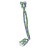 4xa1C 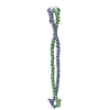 4xa3C  4xa6C 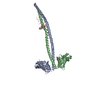 1ik9S C: citing same article ( S: Starting model for refinement |
|---|---|
| Similar structure data |

-Links
 PDBj
PDBj


- Assembly
| Deposited unit | 
| ||||||||
|---|---|---|---|---|---|---|---|---|---|
| 1 |
| ||||||||
| Unit cell |
|
-Components
| #1: Protein | Mass: 23810.619 Da / Num. of mol.: 2 Fragment: UNP Q13426 residues 2-147,UNP P12883 residues 1551-1609 Source method: isolated from a genetically manipulated source Source: (gene. exp.) Homo sapiens (human) / Gene: XRCC4, MYH7, MYHCB / Plasmid: pET28 / Production host:  #2: Water | ChemComp-HOH / |  Mass: 18.015 Da / Num. of mol.: 38 / Source method: isolated from a natural source / Formula: H2O Mass: 18.015 Da / Num. of mol.: 38 / Source method: isolated from a natural source / Formula: H2O |
|---|
-Experimental details
-Experiment
| Experiment | Method: X-RAY DIFFRACTION / Number of used crystals: 1 |
|---|
- Sample preparation
| Crystal | Density Matthews: 2.65 Å3/Da / Density % sol: 54 % |
|---|---|
| Crystal grow | Temperature: 298 K / Method: vapor diffusion, hanging drop / pH: 8.5 Details: 30% (w/v) polyethylene glycol 1500, 250 mM tetramethylammonium chloride, 100 mM 3-[4-(2-Hydroxyethyl)-1-piperazinyl]propanesulfonic acid (HEPPS) |
-Data collection
| Diffraction | Mean temperature: 100 K |
|---|---|
| Diffraction source | Source: SYNCHROTRON / Site: APS / Beamline: 19-ID / Wavelength: 0.9794 Å |
| Detector | Type: ADSC QUANTUM 315r / Detector: CCD / Date: Aug 4, 2012 |
| Radiation | Monochromator: Rosenbaum-Rock double-crystal monochromator: Water cooled; sagitally focusing 2nd crystal, Rosenbaum-Rock vertical focusing mirror Protocol: SINGLE WAVELENGTH / Monochromatic (M) / Laue (L): M / Scattering type: x-ray |
| Radiation wavelength | Wavelength: 0.9794 Å / Relative weight: 1 |
| Reflection | Resolution: 2.327→50 Å / Num. obs: 22289 / % possible obs: 99 % / Redundancy: 7.5 % / Biso Wilson estimate: 38 Å2 / Rmerge(I) obs: 0.06 / Net I/av σ(I): 32.9 / Net I/σ(I): 23.4 |
| Reflection shell | Resolution: 2.327→2.37 Å / Redundancy: 6.5 % / Rmerge(I) obs: 0.31 / Mean I/σ(I) obs: 8.5 / % possible all: 99.9 |
- Processing
| Software |
| |||||||||||||||||||||||||||||||||||||||||||||||||||||||||||||||
|---|---|---|---|---|---|---|---|---|---|---|---|---|---|---|---|---|---|---|---|---|---|---|---|---|---|---|---|---|---|---|---|---|---|---|---|---|---|---|---|---|---|---|---|---|---|---|---|---|---|---|---|---|---|---|---|---|---|---|---|---|---|---|---|---|
| Refinement | Method to determine structure: MOLECULAR REPLACEMENT Starting model: 1IK9 Resolution: 2.327→34.542 Å / SU ML: 0.3 / Cross valid method: FREE R-VALUE / σ(F): 1.34 / Phase error: 27.07 / Stereochemistry target values: ML
| |||||||||||||||||||||||||||||||||||||||||||||||||||||||||||||||
| Solvent computation | Shrinkage radii: 0.9 Å / VDW probe radii: 1.11 Å / Solvent model: FLAT BULK SOLVENT MODEL | |||||||||||||||||||||||||||||||||||||||||||||||||||||||||||||||
| Refinement step | Cycle: LAST / Resolution: 2.327→34.542 Å
| |||||||||||||||||||||||||||||||||||||||||||||||||||||||||||||||
| Refine LS restraints |
| |||||||||||||||||||||||||||||||||||||||||||||||||||||||||||||||
| LS refinement shell |
|
