 Movie
Movie Controller


+ Open data

- Basic information
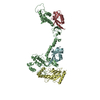
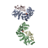
| Entry | Database: PDB / ID: 1ik9 | ||||||
|---|---|---|---|---|---|---|---|
| Title | CRYSTAL STRUCTURE OF A XRCC4-DNA LIGASE IV COMPLEX | ||||||
 Components Components |
| ||||||
 Keywords Keywords | GENE REGULATION/LIGASE / DNA end joining / double-strand break repair / V(D)J recombination / protein-protein complex / coiled coil / GENE REGULATION-LIGASE COMPLEX | ||||||
| Function / homology |  Function and homology information Function and homology informationFHA domain binding / positive regulation of chromosome organization / DN2 thymocyte differentiation / positive regulation of ligase activity / DNA ligase IV complex / DNA ligase activity / DNA double-strand break attachment to nuclear envelope / T cell receptor V(D)J recombination / DNA ligase (ATP) / pro-B cell differentiation ...FHA domain binding / positive regulation of chromosome organization / DN2 thymocyte differentiation / positive regulation of ligase activity / DNA ligase IV complex / DNA ligase activity / DNA double-strand break attachment to nuclear envelope / T cell receptor V(D)J recombination / DNA ligase (ATP) / pro-B cell differentiation / DNA ligase (ATP) activity / DNA-dependent protein kinase-DNA ligase 4 complex / immunoglobulin V(D)J recombination / nonhomologous end joining complex / nucleotide-excision repair, DNA gap filling / single strand break repair / V(D)J recombination / double-strand break repair via classical nonhomologous end joining / isotype switching / protein localization to site of double-strand break / positive regulation of neurogenesis / cellular response to lithium ion / DNA biosynthetic process / 2-LTR circle formation / AMP binding / ligase activity / somatic stem cell population maintenance / chromosome organization / response to X-ray / SUMOylation of DNA damage response and repair proteins / condensed chromosome / neurogenesis / stem cell proliferation / cellular response to ionizing radiation / response to gamma radiation / central nervous system development / Nonhomologous End-Joining (NHEJ) / establishment of integrated proviral latency / base-excision repair / double-strand break repair via nonhomologous end joining / positive regulation of fibroblast proliferation / T cell differentiation in thymus / double-strand break repair / site of double-strand break / neuron apoptotic process / fibroblast proliferation / in utero embryonic development / negative regulation of neuron apoptotic process / protein-macromolecule adaptor activity / chromosome, telomeric region / cell population proliferation / cell division / magnesium ion binding / enzyme binding / DNA binding / nucleoplasm / ATP binding / identical protein binding / nucleus / cytosol Similarity search - Function | ||||||
| Biological species |  Homo sapiens (human) Homo sapiens (human) | ||||||
| Method |  X-RAY DIFFRACTION / SYNCHROTRON / SAS / Resolution: 2.3 Å X-RAY DIFFRACTION / SYNCHROTRON / SAS / Resolution: 2.3 Å | ||||||
 Authors Authors | Sibanda, B.L. / Critchlow, S.E. / Begun, J. / Pei, X.Y. / Jackson, S.P. / Blundell, T.L. / Pellegrini, L. | ||||||
 Citation Citation | Journal: Nat.Struct.Biol. / Year: 2001 Title: Crystal structure of an Xrcc4-DNA ligase IV complex. Authors: Sibanda, B.L. / Critchlow, S.E. / Begun, J. / Pei, X.Y. / Jackson, S.P. / Blundell, T.L. / Pellegrini, L. | ||||||
| History |
|
- Structure visualization
| Structure viewer | Molecule: MolmilJmol/JSmol |
|---|
- Downloads & links
-Download
| PDBx/mmCIF format | 1ik9.cif.gz | 103.5 KB | Display | PDBx/mmCIF format |
|---|---|---|---|---|
| PDB format | pdb1ik9.ent.gz | 80 KB | Display | PDB format |
| PDBx/mmJSON format | 1ik9.json.gz | Tree view | PDBx/mmJSON format | |
| Others |  Other downloads Other downloads |
-Validation report
| Arichive directory | https://data.pdbj.org/pub/pdb/validation_reports/ik/1ik9ftp://data.pdbj.org/pub/pdb/validation_reports/ik/1ik9 | HTTPS FTP |
|---|
-Related structure data
| Similar structure data |
|---|


-Links
 PDBj
PDBj


- Assembly
| Deposited unit | 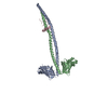
| ||||||||
|---|---|---|---|---|---|---|---|---|---|
| 1 |
| ||||||||
| Unit cell |
|
-Components
| #1: Protein | Mass: 24466.619 Da / Num. of mol.: 2 / Fragment: XRCC4 FRAGMENT, RESIDUES 1-213 / Mutation: C93A,C128A,C130A,C165A Source method: isolated from a genetically manipulated source Source: (gene. exp.) Homo sapiens (human) / Gene: XRCC4 / Plasmid: pET15a / Species (production host): Escherichia coli / Production host:  #2: Protein/peptide | | Mass: 4323.684 Da / Num. of mol.: 1 / Fragment: LINKER CONNECTING BRCT DOMAINS, RESIDUES 748-784 Source method: isolated from a genetically manipulated source Source: (gene. exp.) Homo sapiens (human) / Gene: LIG4 / Plasmid: pTYB3 / Species (production host): Escherichia coli / Production host: #3: Water | ChemComp-HOH / |  Mass: 18.015 Da / Num. of mol.: 224 / Source method: isolated from a natural source / Formula: H2O Mass: 18.015 Da / Num. of mol.: 224 / Source method: isolated from a natural source / Formula: H2O |
|---|
-Experimental details
-Experiment
| Experiment | Method: X-RAY DIFFRACTION / Number of used crystals: 1 |
|---|
- Sample preparation
| Crystal | Density Matthews: 4.3 Å3/Da / Density % sol: 71.5 % | |||||||||||||||||||||||||||||||||||||||||||||||||||||||||||||||
|---|---|---|---|---|---|---|---|---|---|---|---|---|---|---|---|---|---|---|---|---|---|---|---|---|---|---|---|---|---|---|---|---|---|---|---|---|---|---|---|---|---|---|---|---|---|---|---|---|---|---|---|---|---|---|---|---|---|---|---|---|---|---|---|---|
| Crystal grow | Temperature: 291 K / Method: vapor diffusion, hanging drop / pH: 6 Details: PEG6000, MES, xylitol, pH 6.0, VAPOR DIFFUSION, HANGING DROP, temperature 291K | |||||||||||||||||||||||||||||||||||||||||||||||||||||||||||||||
| Crystal grow | *PLUS Details: used seeding | |||||||||||||||||||||||||||||||||||||||||||||||||||||||||||||||
| Components of the solutions | *PLUS
|
-Data collection
| Diffraction | Mean temperature: 100 K |
|---|---|
| Diffraction source | Source: SYNCHROTRON / Site: ESRF  / Beamline: ID14-4 / Wavelength: 0.9795 Å / Beamline: ID14-4 / Wavelength: 0.9795 Å |
| Detector | Type: ADSC QUANTUM 4 / Detector: CCD / Date: Feb 8, 2001 / Details: mirrors |
| Radiation | Monochromator: 111/311 Si crystal / Protocol: SINGLE WAVELENGTH / Monochromatic (M) / Laue (L): M / Scattering type: x-ray |
| Radiation wavelength | Wavelength: 0.9795 Å / Relative weight: 1 |
| Reflection | Resolution: 2.3→33.26 Å / Num. all: 34224 / Num. obs: 34224 / % possible obs: 85.5 % / Observed criterion σ(I): 0 / Redundancy: 2.1 % / Biso Wilson estimate: 56.1 Å2 / Rmerge(I) obs: 0.051 / Rsym value: 0.051 / Net I/σ(I): 17.9 |
| Reflection shell | Resolution: 2.3→2.35 Å / Rmerge(I) obs: 0.229 / Mean I/σ(I) obs: 2.3 / Num. unique all: 795 / Rsym value: 0.229 / % possible all: 44.4 |
| Reflection | *PLUS Num. measured all: 72477 |
| Reflection shell | *PLUS % possible obs: 44.4 % |
- Processing
| Software |
| |||||||||||||||||||||||||
|---|---|---|---|---|---|---|---|---|---|---|---|---|---|---|---|---|---|---|---|---|---|---|---|---|---|---|
| Refinement | Method to determine structure: SAS / Resolution: 2.3→33.26 Å / Isotropic thermal model: overall anisotropic B value / Cross valid method: THROUGHOUT / σ(F): 0 / Stereochemistry target values: Engh & Huber
| |||||||||||||||||||||||||
| Solvent computation | Solvent model: FLAT MODEL / Bsol: 54.1307 Å2 / ksol: 0.340363 e/Å3 | |||||||||||||||||||||||||
| Displacement parameters | Biso mean: 70.4 Å2
| |||||||||||||||||||||||||
| Refine analyze |
| |||||||||||||||||||||||||
| Refinement step | Cycle: LAST / Resolution: 2.3→33.26 Å
| |||||||||||||||||||||||||
| Refine LS restraints |
| |||||||||||||||||||||||||
| LS refinement shell | Resolution: 2.3→2.38 Å / Rfactor Rfree error: 0.048
| |||||||||||||||||||||||||
| Software | *PLUS Name: CNS / Version: 1 / Classification: refinement | |||||||||||||||||||||||||
| Refinement | *PLUS σ(F): 0 / Rfactor obs: 0.228 | |||||||||||||||||||||||||
| Solvent computation | *PLUS | |||||||||||||||||||||||||
| Displacement parameters | *PLUS Biso mean: 70.4 Å2 | |||||||||||||||||||||||||
| Refine LS restraints | *PLUS
| |||||||||||||||||||||||||
| LS refinement shell | *PLUS Rfactor Rfree: 0.467 / Rfactor Rwork: 0.466 |
