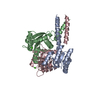
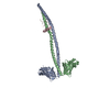
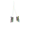
Yeast Spc42 N-terminal coiled-coil fused to PDB: 3K2N
Components
Spindle pole body component SPC42,Sigma-54-dependent transcriptional regulator
Keywords
STRUCTURAL PROTEIN / Yeast Centrosome / Coiled-Coil / Spindle Pole Body
Function / homology
Function and homology information
intermediate layer of spindle pole body / central plaque of spindle pole body / spindle pole body duplication / regulation of microtubule nucleation / structural constituent of cytoskeleton / sequence-specific DNA binding / regulation of DNA-templated transcription / DNA-templated transcription / ATP binding / nucleus Similarity search - Function
Spindle pole body component Spc42 / Spindle pole body component Spc42p / Sigma-54 interaction domain ATP-binding region A signature. / Sigma-54 interaction domain, ATP-binding site 1 / Sigma-54 interaction domain, ATP-binding site 2 / Sigma-54 interaction domain ATP-binding region B signature. / : / AAA+ ATPase lid domain / Sigma-54 interaction domain profile. / Sigma-54 interaction domain ...Spindle pole body component Spc42 / Spindle pole body component Spc42p / Sigma-54 interaction domain ATP-binding region A signature. / Sigma-54 interaction domain, ATP-binding site 1 / Sigma-54 interaction domain, ATP-binding site 2 / Sigma-54 interaction domain ATP-binding region B signature. / : / AAA+ ATPase lid domain / Sigma-54 interaction domain profile. / Sigma-54 interaction domain / RNA polymerase sigma factor 54 interaction domain / DNA binding HTH domain, Fis-type / Bacterial regulatory protein, Fis family / GAF domain / Domain present in phytochromes and cGMP-specific phosphodiesterases. / GAF domain / GAF-like domain superfamily / Homeobox-like domain superfamily / ATPases associated with a variety of cellular activities / AAA+ ATPase domain / P-loop containing nucleoside triphosphate hydrolase Similarity search - Domain/homology
Mass: 18.015 Da / Num. of mol.: 41 / Source method: isolated from a natural source / Formula: H2O
Has protein modification
Y
-
Experimental details
-
Experiment
Experiment
Method: X-RAY DIFFRACTION / Number of used crystals: 1
-
Sample preparation
Crystal
Density Matthews: 3.36 Å3/Da / Density % sol: 63.44 %
Crystal grow
Temperature: 298 K / Method: vapor diffusion, hanging drop / pH: 7 Details: Crystals were grown from a 1:1 mixture of 16 mg/ml protein solution containing 20 mM HEPES pH7.6, 100 mM NaCl and well solution containing 100 mM MOPS pH 7.0, 5% (w/v) PEG4K, 5%(w/v) MPD, and 150 mM NaCitrate.
-
Data collection
Diffraction
Mean temperature: 100 K / Serial crystal experiment: N
In the structure databanks used in Yorodumi, some data are registered as the other names, "COVID-19 virus" and "2019-nCoV". Here are the details of the virus and the list of structure data.
Jan 31, 2019. EMDB accession codes are about to change! (news from PDBe EMDB page)
EMDB accession codes are about to change! (news from PDBe EMDB page)
The allocation of 4 digits for EMDB accession codes will soon come to an end. Whilst these codes will remain in use, new EMDB accession codes will include an additional digit and will expand incrementally as the available range of codes is exhausted. The current 4-digit format prefixed with “EMD-” (i.e. EMD-XXXX) will advance to a 5-digit format (i.e. EMD-XXXXX), and so on. It is currently estimated that the 4-digit codes will be depleted around Spring 2019, at which point the 5-digit format will come into force.
The EM Navigator/Yorodumi systems omit the EMD- prefix.
Related info.:Q: What is EMD? / ID/Accession-code notation in Yorodumi/EM Navigator
Yorodumi is a browser for structure data from EMDB, PDB, SASBDB, etc.
This page is also the successor to EM Navigator detail page, and also detail information page/front-end page for Omokage search.
The word "yorodu" (or yorozu) is an old Japanese word meaning "ten thousand". "mi" (miru) is to see.
Related info.:EMDB / PDB / SASBDB / Comparison of 3 databanks / Yorodumi Search / Aug 31, 2016. New EM Navigator & Yorodumi / Yorodumi Papers / Jmol/JSmol / Function and homology information / Changes in new EM Navigator and Yorodumi
 Movie
Movie Controller


 Open data
 Basic information
 Components
 Keywords
 Function and homology information
 Chlorobaculum tepidum (bacteria)
 X-RAY DIFFRACTION /
  Authors
 United States, 1items
  Citation
 Structure visualization
 Downloads & links
 Other downloads
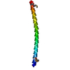


 PDBj
PDBj

 Assembly


 Mass: 118.174 Da / Num. of mol.: 2 / Source method: obtained synthetically / Formula: C6H14O2 / Comment: precipitant*YM
 Mass: 18.015 Da / Num. of mol.: 41 / Source method: isolated from a natural source / Formula: H2O
 Sample preparation
 Processing