 Movie
Movie Controller

[English] 日本語
 Yorodumi
- PDB-3mud: Structure of the Tropomyosin Overlap Complex from Chicken Smooth ... -
+ Open data

- Basic information
| Entry | Database: PDB / ID: 3mud | ||||||
|---|---|---|---|---|---|---|---|
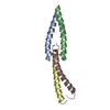
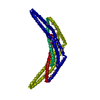
| Title | Structure of the Tropomyosin Overlap Complex from Chicken Smooth Muscle | ||||||
 Components Components |
| ||||||
 Keywords Keywords | CONTRACTILE PROTEIN / tropomysoin / overlap complex / coiled-coils | ||||||
| Function / homology |  Function and homology information Function and homology informationSmooth Muscle Contraction / protein localization to astral microtubule / protein localization to mitotic spindle / cortical microtubule cytoskeleton / FHA domain binding / mitotic spindle astral microtubule end / positive regulation of ligase activity / DNA ligase IV complex / positive regulation of heart rate by epinephrine / muscle thin filament tropomyosin ...Smooth Muscle Contraction / protein localization to astral microtubule / protein localization to mitotic spindle / cortical microtubule cytoskeleton / FHA domain binding / mitotic spindle astral microtubule end / positive regulation of ligase activity / DNA ligase IV complex / positive regulation of heart rate by epinephrine / muscle thin filament tropomyosin / protein localization to microtubule / DNA double-strand break attachment to nuclear envelope / Striated Muscle Contraction / cell projection membrane / microtubule plus-end / DNA-dependent protein kinase-DNA ligase 4 complex / regulation of muscle contraction / immunoglobulin V(D)J recombination / mitotic spindle microtubule / nonhomologous end joining complex / attachment of mitotic spindle microtubules to kinetochore / bleb / ruffle organization / microtubule plus-end binding / microtubule bundle formation / Striated Muscle Contraction / non-motile cilium assembly / protein localization to site of double-strand break / muscle filament sliding / protein localization to centrosome / sarcomere organization / structural constituent of muscle / cellular response to lithium ion / 2-LTR circle formation / ventricular cardiac muscle tissue morphogenesis / mitotic spindle pole / spindle midzone / establishment of mitotic spindle orientation / negative regulation of vascular associated smooth muscle cell migration / regulation of heart contraction / negative regulation of microtubule polymerization / microtubule polymerization / microtubule organizing center / negative regulation of vascular associated smooth muscle cell proliferation / Smooth Muscle Contraction / regulation of microtubule polymerization or depolymerization / response to X-ray / SUMOylation of DNA damage response and repair proteins / positive regulation of microtubule polymerization / cytoplasmic microtubule / cytoskeletal protein binding / spindle assembly / positive regulation of stress fiber assembly / cardiac muscle contraction / stress fiber / cytoskeleton organization / Loss of Nlp from mitotic centrosomes / Loss of proteins required for interphase microtubule organization from the centrosome / Amplification of signal from unattached kinetochores via a MAD2 inhibitory signal / Recruitment of mitotic centrosome proteins and complexes / Recruitment of NuMA to mitotic centrosomes / Anchoring of the basal body to the plasma membrane / Mitotic Prometaphase / positive regulation of cell adhesion / EML4 and NUDC in mitotic spindle formation / AURKA Activation by TPX2 / negative regulation of cell migration / Resolution of Sister Chromatid Cohesion / actin filament organization / protein serine/threonine kinase binding / sarcomere / actin filament / cellular response to reactive oxygen species / wound healing / Nonhomologous End-Joining (NHEJ) / RHO GTPases Activate Formins / base-excision repair / structural constituent of cytoskeleton / double-strand break repair via nonhomologous end joining / ruffle membrane / actin filament binding / The role of GTSE1 in G2/M progression after G2 checkpoint / Separation of Sister Chromatids / intracellular protein localization / Regulation of PLK1 Activity at G2/M Transition / regulation of cell shape / double-strand break repair / cell migration / actin cytoskeleton / site of double-strand break / actin binding / cytoskeleton / microtubule / protein-macromolecule adaptor activity / ciliary basal body / cadherin binding / protein heterodimerization activity / cell division / focal adhesion / centrosome Similarity search - Function | ||||||
| Biological species |  Homo sapiens (human) Homo sapiens (human) | ||||||
| Method |  X-RAY DIFFRACTION / SYNCHROTRON / MOLECULAR REPLACEMENT / Resolution: 2.2 Å X-RAY DIFFRACTION / SYNCHROTRON / MOLECULAR REPLACEMENT / Resolution: 2.2 Å | ||||||
 Authors Authors | Klenchin, V.A. / Frye, J. / Rayment, I. | ||||||
 Citation Citation | Journal: Biochemistry / Year: 2010 Title: Structure of the tropomyosin overlap complex from chicken smooth muscle: insight into the diversity of N-terminal recognition . Authors: Frye, J. / Klenchin, V.A. / Rayment, I. | ||||||
| History |
|
- Structure visualization
| Structure viewer | Molecule: MolmilJmol/JSmol |
|---|
- Downloads & links
-Download
| PDBx/mmCIF format | 3mud.cif.gz | 196.3 KB | Display | PDBx/mmCIF format |
|---|---|---|---|---|
| PDB format | pdb3mud.ent.gz | 157.3 KB | Display | PDB format |
| PDBx/mmJSON format | 3mud.json.gz | Tree view | PDBx/mmJSON format | |
| Others |  Other downloads Other downloads |
-Validation report
| Arichive directory | https://data.pdbj.org/pub/pdb/validation_reports/mu/3mudftp://data.pdbj.org/pub/pdb/validation_reports/mu/3mud | HTTPS FTP |
|---|
-Related structure data
| Related structure data | 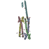 3mtuC 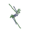 1fu1S C: citing same article ( S: Starting model for refinement |
|---|---|
| Similar structure data |

-Links
 PDBj
PDBj


- Assembly
| Deposited unit | 
| ||||||||
|---|---|---|---|---|---|---|---|---|---|
| 1 |
| ||||||||
| Unit cell |
|
-Components
| #1: Protein | Mass: 19743.168 Da / Num. of mol.: 2 / Mutation: I134T, L249N,I134T Source method: isolated from a genetically manipulated source Source: (gene. exp.) Homo sapiens (human), (gene. exp.) Gene: XRCC4, TPM1 / Plasmid: pET31B / Production host:  #2: Protein | Mass: 8572.687 Da / Num. of mol.: 2 Source method: isolated from a genetically manipulated source Source: (gene. exp.) Homo sapiens (human) / Gene: TPM1, C15orf13, TMSA, MAPRE1 / Plasmid: pET31B / Production host: #3: Chemical | ChemComp-EDO / |   Mass: 62.068 Da / Num. of mol.: 1 / Source method: obtained synthetically / Formula: C2H6O2 Mass: 62.068 Da / Num. of mol.: 1 / Source method: obtained synthetically / Formula: C2H6O2#4: Chemical | ChemComp-SO4 / |   Mass: 96.063 Da / Num. of mol.: 1 / Source method: obtained synthetically / Formula: SO4 Mass: 96.063 Da / Num. of mol.: 1 / Source method: obtained synthetically / Formula: SO4#5: Water | ChemComp-HOH / |  Mass: 18.015 Da / Num. of mol.: 398 / Source method: isolated from a natural source / Formula: H2O Mass: 18.015 Da / Num. of mol.: 398 / Source method: isolated from a natural source / Formula: H2O |
|---|
-Experimental details
-Experiment
| Experiment | Method: X-RAY DIFFRACTION / Number of used crystals: 1 |
|---|
- Sample preparation
| Crystal | Density Matthews: 3.35 Å3/Da / Density % sol: 63.33 % |
|---|---|
| Crystal grow | Temperature: 293 K / Method: vapor diffusion, hanging drop / pH: 6.5 Details: 18% MePEG 2000, 100 mM Bis-Tris, 120 mM MgSO4 , pH 6.5, VAPOR DIFFUSION, HANGING DROP, temperature 293K |
-Data collection
| Diffraction | Mean temperature: 100 K |
|---|---|
| Diffraction source | Source: SYNCHROTRON / Site: APS  / Beamline: 19-BM / Wavelength: 0.9793 Å / Beamline: 19-BM / Wavelength: 0.9793 Å |
| Detector | Type: SBC-3 / Detector: CCD / Date: Aug 6, 2009 |
| Radiation | Monochromator: Rosenbaum-Rock double-crystal / Protocol: SINGLE WAVELENGTH / Monochromatic (M) / Laue (L): M / Scattering type: x-ray |
| Radiation wavelength | Wavelength: 0.9793 Å / Relative weight: 1 |
| Reflection | Resolution: 2.2→30 Å / Num. all: 39926 / Num. obs: 39647 / % possible obs: 99.3 % / Observed criterion σ(F): 0 / Observed criterion σ(I): -3 / Redundancy: 18.6 % / Rmerge(I) obs: 0.057 / Net I/σ(I): 49.6 |
| Reflection shell | Resolution: 2.2→2.24 Å / Redundancy: 17.9 % / Mean I/σ(I) obs: 5.8 / % possible all: 99.2 |
- Processing
| Software |
| |||||||||||||||||||||||||||||||||||||||||||||||||||||||||||||||||||||||||||||||||||||||||||||||||||||||||||||||||||||||||||||||||||||||||||||||||||||||||||||||||||||||||||||||
|---|---|---|---|---|---|---|---|---|---|---|---|---|---|---|---|---|---|---|---|---|---|---|---|---|---|---|---|---|---|---|---|---|---|---|---|---|---|---|---|---|---|---|---|---|---|---|---|---|---|---|---|---|---|---|---|---|---|---|---|---|---|---|---|---|---|---|---|---|---|---|---|---|---|---|---|---|---|---|---|---|---|---|---|---|---|---|---|---|---|---|---|---|---|---|---|---|---|---|---|---|---|---|---|---|---|---|---|---|---|---|---|---|---|---|---|---|---|---|---|---|---|---|---|---|---|---|---|---|---|---|---|---|---|---|---|---|---|---|---|---|---|---|---|---|---|---|---|---|---|---|---|---|---|---|---|---|---|---|---|---|---|---|---|---|---|---|---|---|---|---|---|---|---|---|---|---|
| Refinement | Method to determine structure: MOLECULAR REPLACEMENT Starting model: PDB ENTRY 1FU1 Resolution: 2.2→30 Å / Cor.coef. Fo:Fc: 0.947 / Cor.coef. Fo:Fc free: 0.924 / SU B: 9.66 / SU ML: 0.112 / Cross valid method: THROUGHOUT / ESU R: 0.193 / ESU R Free: 0.18 / Stereochemistry target values: MAXIMUM LIKELIHOOD / Details: HYDROGENS HAVE BEEN ADDED IN THE RIDING POSITIONS
| |||||||||||||||||||||||||||||||||||||||||||||||||||||||||||||||||||||||||||||||||||||||||||||||||||||||||||||||||||||||||||||||||||||||||||||||||||||||||||||||||||||||||||||||
| Solvent computation | Ion probe radii: 0.8 Å / Shrinkage radii: 0.8 Å / VDW probe radii: 1.4 Å / Solvent model: MASK | |||||||||||||||||||||||||||||||||||||||||||||||||||||||||||||||||||||||||||||||||||||||||||||||||||||||||||||||||||||||||||||||||||||||||||||||||||||||||||||||||||||||||||||||
| Displacement parameters | Biso mean: 49.235 Å2
| |||||||||||||||||||||||||||||||||||||||||||||||||||||||||||||||||||||||||||||||||||||||||||||||||||||||||||||||||||||||||||||||||||||||||||||||||||||||||||||||||||||||||||||||
| Refinement step | Cycle: LAST / Resolution: 2.2→30 Å
| |||||||||||||||||||||||||||||||||||||||||||||||||||||||||||||||||||||||||||||||||||||||||||||||||||||||||||||||||||||||||||||||||||||||||||||||||||||||||||||||||||||||||||||||
| Refine LS restraints |
| |||||||||||||||||||||||||||||||||||||||||||||||||||||||||||||||||||||||||||||||||||||||||||||||||||||||||||||||||||||||||||||||||||||||||||||||||||||||||||||||||||||||||||||||
| LS refinement shell | Resolution: 2.2→2.257 Å / Total num. of bins used: 20
| |||||||||||||||||||||||||||||||||||||||||||||||||||||||||||||||||||||||||||||||||||||||||||||||||||||||||||||||||||||||||||||||||||||||||||||||||||||||||||||||||||||||||||||||
| Refinement TLS params. | Method: refined / Refine-ID: X-RAY DIFFRACTION
| |||||||||||||||||||||||||||||||||||||||||||||||||||||||||||||||||||||||||||||||||||||||||||||||||||||||||||||||||||||||||||||||||||||||||||||||||||||||||||||||||||||||||||||||
| Refinement TLS group |
|
