Tropomyosin alpha-1 chain,Microtubule-associated protein RP/EB family member 1
Keywords
CONTRACTILE PROTEIN / tropomysoin / overlap complex / coiled-coils
Function / homology
Function and homology information
viral scaffold / Smooth Muscle Contraction / protein localization to astral microtubule / protein localization to mitotic spindle / cortical microtubule cytoskeleton / mitotic spindle astral microtubule end / protein localization to microtubule / Striated Muscle Contraction / cell projection membrane / microtubule plus-end ...viral scaffold / Smooth Muscle Contraction / protein localization to astral microtubule / protein localization to mitotic spindle / cortical microtubule cytoskeleton / mitotic spindle astral microtubule end / protein localization to microtubule / Striated Muscle Contraction / cell projection membrane / microtubule plus-end / mitotic spindle microtubule / attachment of mitotic spindle microtubules to kinetochore / microtubule plus-end binding / microtubule bundle formation / non-motile cilium assembly / protein localization to centrosome / virion assembly / mitotic spindle pole / spindle midzone / establishment of mitotic spindle orientation / negative regulation of microtubule polymerization / microtubule polymerization / microtubule organizing center / regulation of microtubule polymerization or depolymerization / positive regulation of microtubule polymerization / cytoplasmic microtubule / cytoskeletal protein binding / spindle assembly / cardiac muscle contraction / Loss of Nlp from mitotic centrosomes / Loss of proteins required for interphase microtubule organization from the centrosome / Amplification of signal from unattached kinetochores via a MAD2 inhibitory signal / Recruitment of mitotic centrosome proteins and complexes / Recruitment of NuMA to mitotic centrosomes / Anchoring of the basal body to the plasma membrane / Mitotic Prometaphase / EML4 and NUDC in mitotic spindle formation / AURKA Activation by TPX2 / Resolution of Sister Chromatid Cohesion / actin filament organization / protein serine/threonine kinase binding / actin filament / RHO GTPases Activate Formins / actin filament binding / The role of GTSE1 in G2/M progression after G2 checkpoint / Separation of Sister Chromatids / intracellular protein localization / Regulation of PLK1 Activity at G2/M Transition / cell migration / actin cytoskeleton / microtubule / ciliary basal body / cadherin binding / protein heterodimerization activity / cell division / focal adhesion / centrosome / Golgi apparatus / protein homodimerization activity / DNA binding / RNA binding / identical protein binding / plasma membrane / cytosol Similarity search - Function
Single alpha-helices involved in coiled-coils or other helix-helix interfaces - #400 / Bacteriophage phi-29 scaffolding protein Gp7 / Capsid assembly scaffolding protein Gp7 / Phi29 scaffolding protein / Single alpha-helices involved in coiled-coils or other helix-helix interfaces - #1430 / EB1, C-terminal / Microtubule-associated protein RP/EB / EB1, C-terminal domain superfamily / EB1-C terminal (EB1-C) domain profile. / EB1-like C-terminal motif ...Single alpha-helices involved in coiled-coils or other helix-helix interfaces - #400 / Bacteriophage phi-29 scaffolding protein Gp7 / Capsid assembly scaffolding protein Gp7 / Phi29 scaffolding protein / Single alpha-helices involved in coiled-coils or other helix-helix interfaces - #1430 / EB1, C-terminal / Microtubule-associated protein RP/EB / EB1, C-terminal domain superfamily / EB1-C terminal (EB1-C) domain profile. / EB1-like C-terminal motif / Tropomyosins signature. / Tropomyosin / Tropomyosin / Calponin homology (CH) domain / Calponin homology domain / CH domain superfamily / Calponin homology (CH) domain profile. / Single alpha-helices involved in coiled-coils or other helix-helix interfaces / Up-down Bundle / Mainly Alpha Similarity search - Domain/homology
ETHANOL / Tropomyosin alpha-1 chain / Capsid assembly scaffolding protein / Microtubule-associated protein RP/EB family member 1 Similarity search - Component
Biological species
Gallus gallus (chicken) Homo sapiens (human) Bacillus phage phi29 (virus)
Method
X-RAY DIFFRACTION / SYNCHROTRON / SAD / Resolution: 2.1 Å
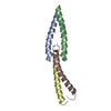
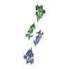
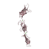
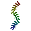
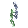
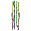
A: Tropomyosin alpha-1 chain,Microtubule-associated protein RP/EB family member 1 B: Tropomyosin alpha-1 chain,Microtubule-associated protein RP/EB family member 1 C: Tropomyosin alpha-1 chain,Microtubule-associated protein RP/EB family member 1 D: Tropomyosin alpha-1 chain,Microtubule-associated protein RP/EB family member 1 E: Capsid assembly scaffolding protein,Tropomyosin alpha-1 chain F: Capsid assembly scaffolding protein,Tropomyosin alpha-1 chain hetero molecules
A: Tropomyosin alpha-1 chain,Microtubule-associated protein RP/EB family member 1 B: Tropomyosin alpha-1 chain,Microtubule-associated protein RP/EB family member 1 C: Tropomyosin alpha-1 chain,Microtubule-associated protein RP/EB family member 1 D: Tropomyosin alpha-1 chain,Microtubule-associated protein RP/EB family member 1 F: Capsid assembly scaffolding protein,Tropomyosin alpha-1 chain hetero molecules
Tropomyosinalpha-1chain,Microtubule-associatedproteinRP/EBfamilymember1 / Alpha-tropomyosin / Tropomyosin-1 / APC-binding protein EB1 / End-binding protein 1 / EB1
Mass: 8713.371 Da / Num. of mol.: 4 Fragment: Fusion protein of residues 1-29 of chicken smooth muscle tropomyosin and residues 215-257 of human EB1 protein,Fusion protein of residues 1-29 of chicken smooth muscle tropomyosin and ...Fragment: Fusion protein of residues 1-29 of chicken smooth muscle tropomyosin and residues 215-257 of human EB1 protein,Fusion protein of residues 1-29 of chicken smooth muscle tropomyosin and residues 215-257 of human EB1 protein Source method: isolated from a genetically manipulated source Source: (gene. exp.) Gallus gallus (chicken), (gene. exp.) Homo sapiens (human) Gene: TPM1, MAPRE1 / Plasmid: pET31B / Production host: Escherichia coli (E. coli) / Strain (production host): BL21(DE3)/pRIL / References: UniProt: P04268, UniProt: Q15691
#2: Protein
Capsidassemblyscaffoldingprotein,Tropomyosinalpha-1chain / Gene product 7 / gp7 / Head morphogenesis protein / Protein p7 / Scaffold protein / Alpha- ...Gene product 7 / gp7 / Head morphogenesis protein / Protein p7 / Scaffold protein / Alpha-tropomyosin / Tropomyosin-1
Mass: 8956.418 Da / Num. of mol.: 2 Fragment: Fusion protein of residues 2-45 of phage phi29 Gp7 protein and residues 256-284 of chicken smooth muscle tropomyosin,Fusion protein of residues 2-45 of phage phi29 Gp7 protein and residues ...Fragment: Fusion protein of residues 2-45 of phage phi29 Gp7 protein and residues 256-284 of chicken smooth muscle tropomyosin,Fusion protein of residues 2-45 of phage phi29 Gp7 protein and residues 256-284 of chicken smooth muscle tropomyosin Source method: isolated from a genetically manipulated source Source: (gene. exp.) Bacillus phage phi29 (virus), (gene. exp.) Gallus gallus (chicken) Plasmid: pET31B / Gene: TPM1 / Production host: Escherichia coli (E. coli) / Strain (production host): BL21(DE3)/pRIL / References: UniProt: P13848, UniProt: P04268
Monochromator: Rosenbaum-Rock double-crystal / Protocol: SINGLE WAVELENGTH / Monochromatic (M) / Laue (L): M / Scattering type: x-ray
Radiation wavelength
Wavelength: 0.9793 Å / Relative weight: 1
Reflection
Resolution: 2.1→30 Å / Num. all: 33163 / Num. obs: 33097 / % possible obs: 99.8 % / Redundancy: 11.8 % / Rmerge(I) obs: 0.104 / Net I/σ(I): 23
Reflection shell
Resolution: 2.1→2.14 Å / Redundancy: 11.1 % / Mean I/σ(I) obs: 3.6 / % possible all: 99.1
-
Processing
Software
Name
Version
Classification
ADSC
Quantum
datacollection
MLPHARE
phasing
REFMAC
5.5.0088
refinement
HKL-3000
datareduction
HKL-2000
datascaling
Refinement
Method to determine structure: SAD / Resolution: 2.1→29.87 Å / Cor.coef. Fo:Fc: 0.94 / Cor.coef. Fo:Fc free: 0.911 / SU B: 8.393 / SU ML: 0.1 / Cross valid method: THROUGHOUT / ESU R: 0.206 / ESU R Free: 0.178 / Stereochemistry target values: MAXIMUM LIKELIHOOD / Details: HYDROGENS HAVE BEEN ADDED IN THE RIDING POSITIONS
Rfactor
Num. reflection
% reflection
Selection details
Rfree
0.24113
1666
5 %
RANDOM
Rwork
0.20096
-
-
-
obs
0.20298
31424
99.64 %
-
all
-
31538
-
-
Solvent computation
Ion probe radii: 0.8 Å / Shrinkage radii: 0.8 Å / VDW probe radii: 1.25 Å / Solvent model: MASK
Displacement parameters
Biso mean: 41.193 Å2
Baniso -1
Baniso -2
Baniso -3
1-
1.44 Å2
0 Å2
0 Å2
2-
-
-0.91 Å2
0 Å2
3-
-
-
-0.52 Å2
Refinement step
Cycle: LAST / Resolution: 2.1→29.87 Å
Protein
Nucleic acid
Ligand
Solvent
Total
Num. atoms
3320
0
59
347
3726
Refine LS restraints
Refine-ID
Type
Dev ideal
Dev ideal target
Number
X-RAY DIFFRACTION
r_bond_refined_d
0.01
0.022
3438
X-RAY DIFFRACTION
r_bond_other_d
X-RAY DIFFRACTION
r_angle_refined_deg
1.069
1.987
4586
X-RAY DIFFRACTION
r_angle_other_deg
X-RAY DIFFRACTION
r_dihedral_angle_1_deg
3.956
5
414
X-RAY DIFFRACTION
r_dihedral_angle_2_deg
40.068
26.888
196
X-RAY DIFFRACTION
r_dihedral_angle_3_deg
15.676
15
689
X-RAY DIFFRACTION
r_dihedral_angle_4_deg
21.251
15
16
X-RAY DIFFRACTION
r_chiral_restr
0.068
0.2
501
X-RAY DIFFRACTION
r_gen_planes_refined
0.004
0.02
2569
X-RAY DIFFRACTION
r_gen_planes_other
X-RAY DIFFRACTION
r_nbd_refined
X-RAY DIFFRACTION
r_nbd_other
X-RAY DIFFRACTION
r_nbtor_refined
X-RAY DIFFRACTION
r_nbtor_other
X-RAY DIFFRACTION
r_xyhbond_nbd_refined
X-RAY DIFFRACTION
r_xyhbond_nbd_other
X-RAY DIFFRACTION
r_metal_ion_refined
X-RAY DIFFRACTION
r_metal_ion_other
X-RAY DIFFRACTION
r_symmetry_vdw_refined
X-RAY DIFFRACTION
r_symmetry_vdw_other
X-RAY DIFFRACTION
r_symmetry_hbond_refined
X-RAY DIFFRACTION
r_symmetry_hbond_other
X-RAY DIFFRACTION
r_symmetry_metal_ion_refined
X-RAY DIFFRACTION
r_symmetry_metal_ion_other
X-RAY DIFFRACTION
r_mcbond_it
2.562
3
2071
X-RAY DIFFRACTION
r_mcbond_other
X-RAY DIFFRACTION
r_mcangle_it
4.922
50
3314
X-RAY DIFFRACTION
r_scbond_it
6.256
5
1367
X-RAY DIFFRACTION
r_scangle_it
9.817
70
1267
X-RAY DIFFRACTION
r_rigid_bond_restr
X-RAY DIFFRACTION
r_sphericity_free
X-RAY DIFFRACTION
r_sphericity_bonded
LS refinement shell
Resolution: 2.098→2.152 Å / Total num. of bins used: 20
Rfactor
Num. reflection
% reflection
Rfree
0.238
118
-
Rwork
0.202
2237
-
obs
-
-
97.64 %
Refinement TLS params.
Method: refined / Refine-ID: X-RAY DIFFRACTION
ID
L11 (°2)
L12 (°2)
L13 (°2)
L22 (°2)
L23 (°2)
L33 (°2)
S11 (Å °)
S12 (Å °)
S13 (Å °)
S21 (Å °)
S22 (Å °)
S23 (Å °)
S31 (Å °)
S32 (Å °)
S33 (Å °)
T11 (Å2)
T12 (Å2)
T13 (Å2)
T22 (Å2)
T23 (Å2)
T33 (Å2)
Origin x (Å)
Origin y (Å)
Origin z (Å)
1
0.1312
-0.258
0.0037
4.905
-0.5298
0.1722
0.0749
0.0478
0.0203
0.1504
-0.0624
0.1864
-0.0109
-0.0249
-0.0125
0.0643
0.0144
0.0281
0.1053
-0.0002
0.0319
11.547
84.504
42.11
2
0.0677
0.3015
0.0791
7.7216
1.1808
0.3238
0.0866
0.001
-0.0121
-0.0047
-0.1406
-0.0915
0.0207
-0.0776
0.054
0.1064
0.0284
-0.0201
0.1315
-0.0051
0.0413
12.049
81.797
44.038
3
0.1217
-0.5689
-0.3274
8.1029
2.349
1.0128
0.0419
-0.017
-0.004
-0.0435
-0.0311
-0.0903
-0.0769
0.0049
-0.0108
0.0628
-0.0093
-0.0318
0.1243
-0.0135
0.0216
12.153
64.556
21.278
4
0.0125
0.1261
-0.0099
4.7034
-0.8349
0.8709
0.0257
0.013
-0.0011
-0.1257
-0.0269
0.0928
-0.0456
-0.0298
0.0012
0.0579
0.0277
-0.0078
0.1279
0.0015
0.0337
9.893
64.489
22.429
5
0.2618
-0.7791
-0.1042
11.6682
0.0689
0.5496
-0.0613
-0.0265
-0.0489
-1.3548
0.109
-0.1243
0.2441
0.0426
-0.0477
0.3321
0.0101
0.0251
0.0816
-0.0216
0.0757
22.827
13.288
32.906
6
6.8174
-12.1873
-2.9096
21.9186
5.2808
1.9678
-0.2567
-0.0892
0.1988
0.6526
0.1386
-0.4463
0.5622
-0.1954
0.1181
0.4397
-0.1499
-0.0239
0.0828
-0.0485
0.1997
23.506
12.93
38.249
Refinement TLS group
ID
Refine-ID
Refine TLS-ID
Auth asym-ID
Auth seq-ID
1
X-RAY DIFFRACTION
1
A
-2 - 257
2
X-RAY DIFFRACTION
2
B
-2 - 257
3
X-RAY DIFFRACTION
3
C
-2 - 257
4
X-RAY DIFFRACTION
4
D
-2 - 257
5
X-RAY DIFFRACTION
5
E
1 - 284
6
X-RAY DIFFRACTION
6
F
1 - 284
+
About Yorodumi
-
News
-
Feb 9, 2022. New format data for meta-information of EMDB entries
New format data for meta-information of EMDB entries
Version 3 of the EMDB header file is now the official format.
The previous official version 1.9 will be removed from the archive.
In the structure databanks used in Yorodumi, some data are registered as the other names, "COVID-19 virus" and "2019-nCoV". Here are the details of the virus and the list of structure data.
Jan 31, 2019. EMDB accession codes are about to change! (news from PDBe EMDB page)
EMDB accession codes are about to change! (news from PDBe EMDB page)
The allocation of 4 digits for EMDB accession codes will soon come to an end. Whilst these codes will remain in use, new EMDB accession codes will include an additional digit and will expand incrementally as the available range of codes is exhausted. The current 4-digit format prefixed with “EMD-” (i.e. EMD-XXXX) will advance to a 5-digit format (i.e. EMD-XXXXX), and so on. It is currently estimated that the 4-digit codes will be depleted around Spring 2019, at which point the 5-digit format will come into force.
The EM Navigator/Yorodumi systems omit the EMD- prefix.
Related info.:Q: What is EMD? / ID/Accession-code notation in Yorodumi/EM Navigator
Yorodumi is a browser for structure data from EMDB, PDB, SASBDB, etc.
This page is also the successor to EM Navigator detail page, and also detail information page/front-end page for Omokage search.
The word "yorodu" (or yorozu) is an old Japanese word meaning "ten thousand". "mi" (miru) is to see.
Related info.:EMDB / PDB / SASBDB / Comparison of 3 databanks / Yorodumi Search / Aug 31, 2016. New EM Navigator & Yorodumi / Yorodumi Papers / Jmol/JSmol / Function and homology information / Changes in new EM Navigator and Yorodumi
 Movie
Movie Controller

 Yorodumi
 Open data
 Basic information
 Components
 Keywords
 Function and homology information
 Homo sapiens (human)
 Bacillus phage phi29 (virus)
 X-RAY DIFFRACTION /
  Authors
 Citation
 Structure visualization
 Downloads & links
 Other downloads
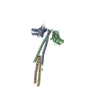

 PDBj
PDBj


 Assembly
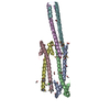


 Mass: 35.453 Da / Num. of mol.: 2 / Source method: obtained synthetically / Formula: Cl
 Mass: 62.068 Da / Num. of mol.: 9 / Source method: obtained synthetically / Formula: C2H6O2
 Mass: 46.068 Da / Num. of mol.: 7 / Source method: obtained synthetically / Formula: C2H6O
 Sample preparation
 / Beamline: 19-BM / Wavelength: 0.9793 Å
 Processing