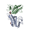
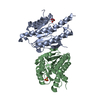
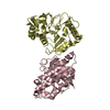
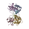
Entry Database : PDB / ID : 5c67Title Human Mesotrypsin in complex with amyloid precursor protein inhibitor variant APPI-M17G/I18F/F34V Amyloid beta A4 protein Trypsin-3 Keywords / / / / Function / homology Function Domain/homology Component
/ / / / / / / / / / / / / / / / / / / / / / / / / / / / / / / / / / / / / / / / / / / / / / / / / / / / / / / / / / / / / / / / / / / / / / / / / / / / / / / / / / / / / / / / / / / / / / / / / / / / / / / / / / / / / / / / / / / / / / / / / / / / / / / / / / / / / / / / / / / / / / / / / / / / / / / / / / / / / / / / / / / / / Biological species Homo sapiens (human)Method / / / Resolution : 1.83 Å Authors Kayode, O. / Sankaran, B. / Radisky, E.S. Funding support Organization Grant number Country National Institutes of Health/National Cancer Institute (NIH/NCI) R01CA154387
Journal : Biochem.J. / Year : 2016Title : Combinatorial protein engineering of proteolytically resistant mesotrypsin inhibitors as candidates for cancer therapy.Authors : Cohen, I. / Kayode, O. / Hockla, A. / Sankaran, B. / Radisky, D.C. / Radisky, E.S. / Papo, N. History Deposition Jun 22, 2015 Deposition site / Processing site Revision 1.0 May 4, 2016 Provider / Type Revision 1.1 May 18, 2016 Group / Source and taxonomyRevision 1.2 Jun 1, 2016 Group Revision 1.3 Sep 6, 2017 Group / Database references / Derived calculationsCategory / pdbx_audit_support / pdbx_struct_oper_listItem / _pdbx_audit_support.funding_organization / _pdbx_struct_oper_list.symmetry_operationRevision 1.4 Dec 4, 2019 Group / Derived calculationsCategory / pdbx_struct_assembly_gen / pdbx_struct_assembly_propItem / _pdbx_struct_assembly_gen.asym_id_list / _pdbx_struct_assembly_prop.valueRevision 1.5 Sep 27, 2023 Group / Database references / Refinement descriptionCategory chem_comp_atom / chem_comp_bond ... chem_comp_atom / chem_comp_bond / database_2 / pdbx_initial_refinement_model Item / _database_2.pdbx_database_accessionRevision 1.6 Oct 23, 2024 Group / Category / pdbx_modification_feature
Show all Show less
 Movie
Movie Controller

 Yorodumi
 Open data
 Basic information
 Components
 Keywords
 Function and homology information
 Homo sapiens (human)
 X-RAY DIFFRACTION /
  Authors
 United States, 1items
  Citation
 Structure visualization
 Downloads & links
 Other downloads


 PDBj
PDBj


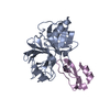
 Assembly
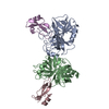


 Komagataella pastoris GS115 (fungus) / Strain (production host): GS115 / References: UniProt: P05067
 Mass: 18.015 Da / Num. of mol.: 87 / Source method: isolated from a natural source / Formula: H2O
 Sample preparation
 Processing