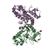
| Deposited unit | A: Uracil-DNA glycosylase
B: Uracil-DNA glycosylase
C: Uracil-DNA glycosylase
D: Uracil-DNA glycosylase
E: Uracil-DNA glycosylase
F: Uracil-DNA glycosylase
G: Uracil-DNA glycosylase
H: Uracil-DNA glycosylase
I: Uracil-DNA glycosylase
J: Uracil-DNA glycosylase
K: Uracil-DNA glycosylase
L: Uracil-DNA glycosylase
hetero molecules
| Theoretical mass | Number of molelcules |
|---|
| Total (without water) | 331,044 | 74 |
|---|
| Polymers | 327,037 | 12 |
|---|
| Non-polymers | 4,007 | 62 |
|---|
| Water | 29,130 | 1617 |
|---|
|
|---|
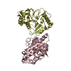
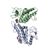
| 1 | A: Uracil-DNA glycosylase
F: Uracil-DNA glycosylase
hetero molecules
| Theoretical mass | Number of molelcules |
|---|
| Total (without water) | 55,320 | 15 |
|---|
| Polymers | 54,506 | 2 |
|---|
| Non-polymers | 813 | 13 |
|---|
| Water | 36 | 2 |
|---|
| Type | Name | Symmetry operation | Number |
|---|
| identity operation | 1_555 | x,y,z | 1 |
|
|---|
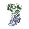
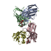
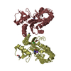
| 2 | B: Uracil-DNA glycosylase
E: Uracil-DNA glycosylase
hetero molecules
| Theoretical mass | Number of molelcules |
|---|
| Total (without water) | 55,625 | 21 |
|---|
| Polymers | 54,506 | 2 |
|---|
| Non-polymers | 1,119 | 19 |
|---|
| Water | 36 | 2 |
|---|
| Type | Name | Symmetry operation | Number |
|---|
| identity operation | 1_555 | x,y,z | 1 |
|
|---|
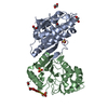
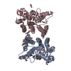
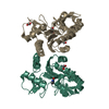
| 3 | C: Uracil-DNA glycosylase
K: Uracil-DNA glycosylase
hetero molecules
| Theoretical mass | Number of molelcules |
|---|
| Total (without water) | 54,902 | 8 |
|---|
| Polymers | 54,506 | 2 |
|---|
| Non-polymers | 396 | 6 |
|---|
| Water | 36 | 2 |
|---|
| Type | Name | Symmetry operation | Number |
|---|
| identity operation | 1_555 | x,y,z | 1 |
|
|---|
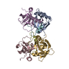
| 4 | D: Uracil-DNA glycosylase
H: Uracil-DNA glycosylase
hetero molecules
| Theoretical mass | Number of molelcules |
|---|
| Total (without water) | 55,121 | 11 |
|---|
| Polymers | 54,506 | 2 |
|---|
| Non-polymers | 615 | 9 |
|---|
| Water | 36 | 2 |
|---|
| Type | Name | Symmetry operation | Number |
|---|
| identity operation | 1_555 | x,y,z | 1 |
|
|---|
| 5 | G: Uracil-DNA glycosylase
J: Uracil-DNA glycosylase
hetero molecules
| Theoretical mass | Number of molelcules |
|---|
| Total (without water) | 55,160 | 12 |
|---|
| Polymers | 54,506 | 2 |
|---|
| Non-polymers | 654 | 10 |
|---|
| Water | 36 | 2 |
|---|
| Type | Name | Symmetry operation | Number |
|---|
| identity operation | 1_555 | x,y,z | 1 |
|
|---|
| 6 | I: Uracil-DNA glycosylase
L: Uracil-DNA glycosylase
hetero molecules
| Theoretical mass | Number of molelcules |
|---|
| Total (without water) | 54,917 | 7 |
|---|
| Polymers | 54,506 | 2 |
|---|
| Non-polymers | 410 | 5 |
|---|
| Water | 36 | 2 |
|---|
| Type | Name | Symmetry operation | Number |
|---|
| identity operation | 1_555 | x,y,z | 1 |
|
|---|
| Unit cell | | Length a, b, c (Å) | 93.470, 114.045, 302.516 |
|---|
| Angle α, β, γ (deg.) | 90.00, 90.00, 90.00 |
|---|
| Int Tables number | 19 |
|---|
| Space group name H-M | P212121 |
|---|
|
|---|
| Noncrystallographic symmetry (NCS) | NCS domain: | ID | Ens-ID | Details (eV) |
|---|
| 1 | 1 | A| 2 | 1 | B| 3 | 1 | C| 4 | 1 | D| 5 | 1 | E| 6 | 1 | F| 7 | 1 | G| 8 | 1 | H| 9 | 1 | I| 10 | 1 | J| 11 | 1 | K| 12 | 1 | L | | | | | | | | | | | |
NCS domain segments: Component-ID: 1 / Ens-ID: 1 / Beg auth comp-ID: MET / Beg label comp-ID: MET / End auth comp-ID: TYR / End label comp-ID: TYR / Refine code: 6 / Auth seq-ID: 1 - 218 / Label seq-ID: 21 - 238 | Dom-ID | Auth asym-ID | Label asym-ID |
|---|
| 1 | AA| 2 | BB| 3 | CC| 4 | DD| 5 | EE| 6 | FF| 7 | GG| 8 | HH| 9 | II| 10 | JJ| 11 | KK| 12 | L| L | | | | | | | | | | | | | | | | | | | | | | | |
|
|---|
 Movie
Movie Controller


 Open data
 Basic information
 Components
 Keywords
 Function and homology information
 Vaccinia virus
 X-RAY DIFFRACTION /
  Authors
 Citation
 Structure visualization
 Downloads & links
 Other downloads


 PDBj
PDBj Assembly
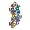


 Mass: 78.133 Da / Num. of mol.: 10 / Source method: obtained synthetically / Formula: C2H6OS / Comment: DMSO, precipitant*YM
 Mass: 39.098 Da / Num. of mol.: 10 / Source method: obtained synthetically / Formula: K
 Mass: 35.453 Da / Num. of mol.: 14 / Source method: obtained synthetically / Formula: Cl
 Mass: 62.068 Da / Num. of mol.: 16 / Source method: obtained synthetically / Formula: C2H6O2
 Mass: 112.087 Da / Num. of mol.: 12 / Source method: obtained synthetically / Formula: C4H4N2O2
 Sample preparation
 / Beamline: 24-ID-C / Wavelength: 1.77 Å
 Processing