Mass: 18.015 Da / Num. of mol.: 800 / Source method: isolated from a natural source / Formula: H2O
Sequence details
Western Reserve (WR109) vaccinia virus strain was used. The closest sequence database entry is: ...Western Reserve (WR109) vaccinia virus strain was used. The closest sequence database entry is: AA089388. Any sequence difference from this entry except for the engineered mutations in the D4 mutants is unintentional
-
Experimental details
-
Experiment
Experiment
Method: X-RAY DIFFRACTION / Number of used crystals: 1
-
Sample preparation
Crystal
Density Matthews: 2.73 Å3/Da / Density % sol: 55.03 %
Crystal grow
Temperature: 277 K / Method: vapor diffusion, hanging drop / pH: 7.5 / Details: 5% PEG3000, 0.1M NaCl, 0.1M Hepes
Monochromator: Double Crystal Si / Protocol: SINGLE WAVELENGTH / Monochromatic (M) / Laue (L): M / Scattering type: x-ray
Radiation wavelength
Wavelength: 0.9762 Å / Relative weight: 1
Reflection
Resolution: 2.3→32.24 Å / Num. obs: 98092 / % possible obs: 100 % / Observed criterion σ(I): 1 / Redundancy: 7.48 % / Biso Wilson estimate: 28.6 Å2 / Rmerge(I) obs: 0.06 / Net I/σ(I): 10.4
Reflection shell
Resolution: 2.3→2.38 Å / Redundancy: 7.48 % / Rmerge(I) obs: 0.253 / Mean I/σ(I) obs: 2.1 / % possible all: 100
-
Processing
Software
Name
Version
Classification
REFMAC
5.8.0131
refinement
HKL-2000
datareduction
SCALEPACK
datascaling
PHASER
phasing
Refinement
Method to determine structure: MOLECULAR REPLACEMENT Starting model: 4DOF_A Resolution: 2.3→32.24 Å / Cor.coef. Fo:Fc: 0.926 / Cor.coef. Fo:Fc free: 0.905 / SU B: 6.613 / SU ML: 0.163 / Data cutoff high absF: 0 / Cross valid method: THROUGHOUT / ESU R: 0.34 / ESU R Free: 0.234 / Details: HYDROGENS HAVE BEEN ADDED IN THE RIDING POSITIONS
Rfactor
Num. reflection
% reflection
Selection details
Rfree
0.24853
4905
5 %
RANDOM
Rwork
0.2152
-
-
-
obs
0.21688
93226
99.91 %
-
Solvent computation
Ion probe radii: 0.8 Å / Shrinkage radii: 0.8 Å / VDW probe radii: 1.2 Å
 Movie
Movie Controller


 Open data
 Basic information
 Components
 Keywords
 Function and homology information
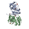
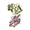
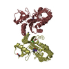
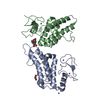
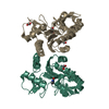
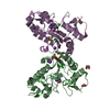
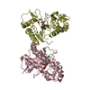
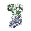
 Vaccinia virus
 X-RAY DIFFRACTION /
  Authors
 United States, 1items
  Citation
 Structure visualization
 Downloads & links
 Other downloads
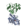


 PDBj
PDBj Assembly
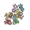


 Mass: 92.094 Da / Num. of mol.: 15 / Source method: obtained synthetically / Formula: C3H8O3
 Mass: 35.453 Da / Num. of mol.: 4 / Source method: obtained synthetically / Formula: Cl
 Mass: 18.015 Da / Num. of mol.: 800 / Source method: isolated from a natural source / Formula: H2O
 Sample preparation
 Processing