- PDB-5c5a: Crystal Structure of HDM2 in complex with Nutlin-3a -
+
データを開く
IDまたはキーワード:
読み込み中...
-
基本情報
登録情報
データベース: PDB / ID: 5c5a
タイトル
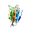
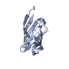
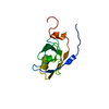
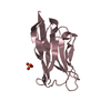
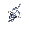
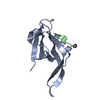
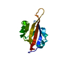
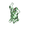
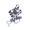
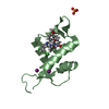
Crystal Structure of HDM2 in complex with Nutlin-3a
要素
E3 ubiquitin-protein ligase Mdm2
キーワード
LIGASE / Drug Design / HDM2 / complex structure
機能・相同性
機能・相同性情報
cellular response to vitamin B1 / response to formaldehyde / response to water-immersion restraint stress / traversing start control point of mitotic cell cycle / negative regulation of intrinsic apoptotic signaling pathway by p53 class mediator / response to ether / fibroblast activation / atrial septum development / regulation of protein catabolic process at postsynapse, modulating synaptic transmission / negative regulation of signal transduction by p53 class mediator ...cellular response to vitamin B1 / response to formaldehyde / response to water-immersion restraint stress / traversing start control point of mitotic cell cycle / negative regulation of intrinsic apoptotic signaling pathway by p53 class mediator / response to ether / fibroblast activation / atrial septum development / regulation of protein catabolic process at postsynapse, modulating synaptic transmission / negative regulation of signal transduction by p53 class mediator / receptor serine/threonine kinase binding / Trafficking of AMPA receptors / peroxisome proliferator activated receptor binding / positive regulation of vascular associated smooth muscle cell migration / negative regulation of protein processing / SUMO transferase activity / response to iron ion / NEDD8 ligase activity / AKT phosphorylates targets in the cytosol / atrioventricular valve morphogenesis / response to steroid hormone / cellular response to peptide hormone stimulus / ventricular septum development / endocardial cushion morphogenesis / positive regulation of muscle cell differentiation / cellular response to alkaloid / SUMOylation of ubiquitinylation proteins / regulation of postsynaptic neurotransmitter receptor internalization / cardiac septum morphogenesis / blood vessel development / ligase activity / Constitutive Signaling by AKT1 E17K in Cancer / regulation of protein catabolic process / negative regulation of DNA damage response, signal transduction by p53 class mediator / response to magnesium ion / SUMOylation of transcription factors / protein sumoylation / cellular response to estrogen stimulus / cellular response to UV-C / blood vessel remodeling / cellular response to actinomycin D / protein localization to nucleus / ribonucleoprotein complex binding / protein autoubiquitination / positive regulation of vascular associated smooth muscle cell proliferation / NPAS4 regulates expression of target genes / transcription repressor complex / positive regulation of mitotic cell cycle / regulation of heart rate / proteolysis involved in protein catabolic process / DNA damage response, signal transduction by p53 class mediator / ubiquitin binding / positive regulation of protein export from nucleus / Stabilization of p53 / response to cocaine / establishment of protein localization / Regulation of RUNX3 expression and activity / cellular response to gamma radiation / protein destabilization / RING-type E3 ubiquitin transferase / cellular response to growth factor stimulus / Oncogene Induced Senescence / Regulation of TP53 Activity through Methylation / centriolar satellite / cellular response to hydrogen peroxide / response to toxic substance / protein polyubiquitination / ubiquitin-protein transferase activity / disordered domain specific binding / p53 binding / endocytic vesicle membrane / ubiquitin protein ligase activity / Signaling by ALK fusions and activated point mutants / Regulation of TP53 Degradation / positive regulation of proteasomal ubiquitin-dependent protein catabolic process / negative regulation of neuron projection development / 5S rRNA binding / protein-containing complex assembly / ubiquitin-dependent protein catabolic process / Oxidative Stress Induced Senescence / cellular response to hypoxia / Regulation of TP53 Activity through Phosphorylation / amyloid fibril formation / proteasome-mediated ubiquitin-dependent protein catabolic process / regulation of cell cycle / postsynaptic density / Ub-specific processing proteases / protein ubiquitination / protein domain specific binding / response to xenobiotic stimulus / response to antibiotic / negative regulation of DNA-templated transcription / apoptotic process / positive regulation of cell population proliferation / ubiquitin protein ligase binding / positive regulation of gene expression / negative regulation of apoptotic process / nucleolus / glutamatergic synapse / enzyme binding 類似検索 - 分子機能
E3 ubiquitin-protein ligase Mdm2 / MDM2, modified RING finger, HC subclass / MDM2 / SWIB/MDM2 domain / p53 negative regulator Mdm2/Mdm4 / SWIB/MDM2 domain / SWIB/MDM2 domain / SWIB/MDM2 domain profile. / SWIB/MDM2 domain superfamily / Zn-finger in Ran binding protein and others ...E3 ubiquitin-protein ligase Mdm2 / MDM2, modified RING finger, HC subclass / MDM2 / SWIB/MDM2 domain / p53 negative regulator Mdm2/Mdm4 / SWIB/MDM2 domain / SWIB/MDM2 domain / SWIB/MDM2 domain profile. / SWIB/MDM2 domain superfamily / Zn-finger in Ran binding protein and others / Zinc finger, C3HC4 type (RING finger) / Zinc finger RanBP2 type profile. / Zinc finger, RanBP2-type superfamily / Zinc finger RanBP2-type signature. / Zinc finger, RanBP2-type / Zinc finger RING-type profile. / Zinc finger, RING-type / Zinc finger, RING/FYVE/PHD-type / Orthogonal Bundle / Mainly Alpha 類似検索 - ドメイン・相同性
 ムービー
ムービー コントローラー


 データを開く
 基本情報
基本情報 要素
要素 キーワード
キーワード 機能・相同性情報
 Homo sapiens (ヒト)
 X線回折 /
  データ登録者
 スイス, 3件
  引用
引用 構造の表示
構造の表示 ダウンロードとリンク
 その他のダウンロード


 PDBj
PDBj


 集合体
集合体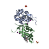


 分子量: 581.490 Da / 分子数: 2 / 由来タイプ: 合成 / 式: C30H30Cl2N4O4
 分子量: 96.063 Da / 分子数: 2 / 由来タイプ: 合成 / 式: SO4
 分子量: 35.453 Da / 分子数: 2 / 由来タイプ: 合成 / 式: Cl
 分子量: 126.904 Da / 分子数: 4 / 由来タイプ: 合成 / 式: I
 試料調製
試料調製 解析
解析