 Movie
Movie Controller


+ Open data

- Basic information
| Entry | Database: PDB / ID: 1rzx | ||||||
|---|---|---|---|---|---|---|---|
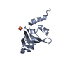
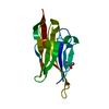
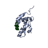
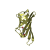
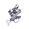
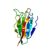
| Title | Crystal Structure of a Par-6 PDZ-peptide Complex | ||||||
 Components Components |
| ||||||
 Keywords Keywords | CELL CYCLE | ||||||
| Function / homology |  Function and homology information Function and homology informationRAC1 GTPase cycle / RHOV GTPase cycle / terminal branching, open tracheal system / establishment or maintenance of neuroblast polarity / branching involved in open tracheal system development / zonula adherens assembly / subapical complex / TGF-beta receptor signaling in EMT (epithelial to mesenchymal transition) / establishment of neuroblast polarity / Asymmetric localization of PCP proteins ...RAC1 GTPase cycle / RHOV GTPase cycle / terminal branching, open tracheal system / establishment or maintenance of neuroblast polarity / branching involved in open tracheal system development / zonula adherens assembly / subapical complex / TGF-beta receptor signaling in EMT (epithelial to mesenchymal transition) / establishment of neuroblast polarity / Asymmetric localization of PCP proteins / RHOU GTPase cycle / muscle cell postsynaptic specialization / establishment or maintenance of polarity of embryonic epithelium / border follicle cell migration / PAR polarity complex / asymmetric neuroblast division / zonula adherens / apical cortex / establishment or maintenance of epithelial cell apical/basal polarity / morphogenesis of a polarized epithelium / positive regulation of smoothened signaling pathway / apical protein localization / positive regulation of filopodium assembly / centrosome cycle / protein kinase inhibitor activity / bicellular tight junction / positive regulation of lamellipodium assembly / synapse assembly / apical part of cell / terminal bouton / cell cortex / apical plasma membrane / plasma membrane Similarity search - Function | ||||||
| Biological species |  | ||||||
| Method |  X-RAY DIFFRACTION / SYNCHROTRON / MOLECULAR REPLACEMENT / Resolution: 2.1 Å X-RAY DIFFRACTION / SYNCHROTRON / MOLECULAR REPLACEMENT / Resolution: 2.1 Å | ||||||
 Authors Authors | Peterson, F.C. / Penkert, R.R. / Volkman, F.B. / Prehoda, K.E. | ||||||
 Citation Citation | Journal: Mol.Cell / Year: 2004 Title: Cdc42 regulates the Par-6 PDZ domain through an allosteric CRIB-PDZ transition. Authors: Peterson, F.C. / Penkert, R.R. / Volkman, B.F. / Prehoda, K.E. | ||||||
| History |
|
- Structure visualization
| Structure viewer | Molecule: MolmilJmol/JSmol |
|---|
- Downloads & links
-Download
| PDBx/mmCIF format | 1rzx.cif.gz | 31.9 KB | Display | PDBx/mmCIF format |
|---|---|---|---|---|
| PDB format | pdb1rzx.ent.gz | 21.5 KB | Display | PDB format |
| PDBx/mmJSON format | 1rzx.json.gz | Tree view | PDBx/mmJSON format | |
| Others |  Other downloads Other downloads |
-Validation report
| Arichive directory | https://data.pdbj.org/pub/pdb/validation_reports/rz/1rzxftp://data.pdbj.org/pub/pdb/validation_reports/rz/1rzx | HTTPS FTP |
|---|
-Related structure data
| Related structure data |  1ry4C  1nf3S S: Starting model for refinement C: citing same article ( |
|---|---|
| Similar structure data |


-Links
 PDBj
PDBj

- Assembly
| Deposited unit | 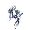
| |||||||||
|---|---|---|---|---|---|---|---|---|---|---|
| 1 |
| |||||||||
| Unit cell |
| |||||||||
| Components on special symmetry positions |
|
-Components
| #1: Protein | Mass: 10497.011 Da / Num. of mol.: 1 Source method: isolated from a genetically manipulated source Source: (gene. exp.)  |
|---|---|
| #2: Protein/peptide | Mass: 700.844 Da / Num. of mol.: 1 / Source method: obtained synthetically Details: This protein was chemically synthesized. The N-terminal has been acetylated (ACE)VKESLV. |
| #3: Water | ChemComp-HOH /  Mass: 18.015 Da / Num. of mol.: 45 / Source method: isolated from a natural source / Formula: H2O Mass: 18.015 Da / Num. of mol.: 45 / Source method: isolated from a natural source / Formula: H2O |
| Has protein modification | Y |
-Experimental details
-Experiment
| Experiment | Method: X-RAY DIFFRACTION / Number of used crystals: 1 |
|---|
- Sample preparation
| Crystal | Density Matthews: 2.85 Å3/Da / Density % sol: 56.85 % | ||||||||||||||||||||||||
|---|---|---|---|---|---|---|---|---|---|---|---|---|---|---|---|---|---|---|---|---|---|---|---|---|---|
| Crystal grow | Temperature: 288 K / Method: vapor diffusion, hanging drop / pH: 7.1 Details: 26% PEG6000, 100 mM HEPES, pH 7.1, VAPOR DIFFUSION, HANGING DROP, temperature 288K | ||||||||||||||||||||||||
| Crystal grow | *PLUS Temperature: 16 ℃ / Method: vapor diffusion, hanging drop | ||||||||||||||||||||||||
| Components of the solutions | *PLUS
|
-Data collection
| Diffraction source | Source: SYNCHROTRON / Site: ALS  / Beamline: 8.2.2 / Wavelength: 1 Å / Beamline: 8.2.2 / Wavelength: 1 Å |
|---|---|
| Radiation | Protocol: SINGLE WAVELENGTH / Monochromatic (M) / Laue (L): M / Scattering type: x-ray |
| Radiation wavelength | Wavelength: 1 Å / Relative weight: 1 |
| Reflection | Resolution: 2.1→30 Å / Num. all: 7402 / Num. obs: 7357 / % possible obs: 99.4 % |
| Reflection shell | Resolution: 2.1→2.18 Å / % possible all: 96 |
| Reflection | *PLUS Rmerge(I) obs: 0.036 |
| Reflection shell | *PLUS % possible obs: 96.1 % / Num. unique obs: 693 / Rmerge(I) obs: 0.122 / Mean I/σ(I) obs: 10.3 |
- Processing
| Software |
| ||||||||||||||||||||
|---|---|---|---|---|---|---|---|---|---|---|---|---|---|---|---|---|---|---|---|---|---|
| Refinement | Method to determine structure: MOLECULAR REPLACEMENT Starting model: 1nf3 Resolution: 2.1→30 Å
| ||||||||||||||||||||
| Refinement step | Cycle: LAST / Resolution: 2.1→30 Å
| ||||||||||||||||||||
| Refinement | *PLUS Rfactor Rfree: 0.26 | ||||||||||||||||||||
| Solvent computation | *PLUS | ||||||||||||||||||||
| Displacement parameters | *PLUS | ||||||||||||||||||||
| Refine LS restraints | *PLUS
|
