 Movie
Movie Controller

[English] 日本語
 Yorodumi
- PDB-5c42: Crystal Structure of HIV-1 Reverse Transcriptase (K101P) Variant ... -
+ Open data

- Basic information
| Entry | Database: PDB / ID: 5c42 | ||||||||||||
|---|---|---|---|---|---|---|---|---|---|---|---|---|---|
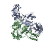
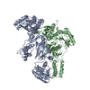
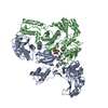
| Title | Crystal Structure of HIV-1 Reverse Transcriptase (K101P) Variant in Complex with 8-(2-(2-(2,4-dioxo-3,4-dihydropyrimidin-1(2H)-yl)ethoxy)phenoxy)indolizine-2-carbonitrile (JLJ555), a non-nucleoside inhibitor | ||||||||||||
 Components Components |
| ||||||||||||
 Keywords Keywords | TRANSFERASE / HYDROLASE/INHIBITOR / HIV / reverse transcriptase / polymerase / non-nucleoside inhibitor / resistance / HYDROLASE-INHIBITOR complex | ||||||||||||
| Function / homology |  Function and homology information Function and homology informationHIV-1 retropepsin / symbiont-mediated activation of host apoptosis / retroviral ribonuclease H / exoribonuclease H / exoribonuclease H activity / host multivesicular body / DNA integration / viral genome integration into host DNA / RNA-directed DNA polymerase / establishment of integrated proviral latency ...HIV-1 retropepsin / symbiont-mediated activation of host apoptosis / retroviral ribonuclease H / exoribonuclease H / exoribonuclease H activity / host multivesicular body / DNA integration / viral genome integration into host DNA / RNA-directed DNA polymerase / establishment of integrated proviral latency / viral penetration into host nucleus / RNA stem-loop binding / RNA-directed DNA polymerase activity / RNA-DNA hybrid ribonuclease activity / Transferases; Transferring phosphorus-containing groups; Nucleotidyltransferases / host cell / viral nucleocapsid / DNA recombination / DNA-directed DNA polymerase / aspartic-type endopeptidase activity / Hydrolases; Acting on ester bonds / DNA-directed DNA polymerase activity / symbiont-mediated suppression of host gene expression / viral translational frameshifting / lipid binding / symbiont entry into host cell / host cell nucleus / host cell plasma membrane / virion membrane / structural molecule activity / proteolysis / DNA binding / zinc ion binding / membrane Similarity search - Function | ||||||||||||
| Biological species |  Human immunodeficiency virus type 1 group M subtype B Human immunodeficiency virus type 1 group M subtype B | ||||||||||||
| Method |  X-RAY DIFFRACTION / SYNCHROTRON / Resolution: 3.5 Å X-RAY DIFFRACTION / SYNCHROTRON / Resolution: 3.5 Å | ||||||||||||
 Authors Authors | Frey, K.M. / Gray, W.T. / Anderson, K.S. | ||||||||||||
| Funding support |  United States, 3items United States, 3items
| ||||||||||||
 Citation Citation | Journal: Acs Med.Chem.Lett. / Year: 2015 Title: Potent Inhibitors Active against HIV Reverse Transcriptase with K101P, a Mutation Conferring Rilpivirine Resistance. Authors: Gray, W.T. / Frey, K.M. / Laskey, S.B. / Mislak, A.C. / Spasov, K.A. / Lee, W.G. / Bollini, M. / Siliciano, R.F. / Jorgensen, W.L. / Anderson, K.S. | ||||||||||||
| History |
|
- Structure visualization
| Structure viewer | Molecule: MolmilJmol/JSmol |
|---|
- Downloads & links
-Download
| PDBx/mmCIF format | 5c42.cif.gz | 210.6 KB | Display | PDBx/mmCIF format |
|---|---|---|---|---|
| PDB format | pdb5c42.ent.gz | 165.3 KB | Display | PDB format |
| PDBx/mmJSON format | 5c42.json.gz | Tree view | PDBx/mmJSON format | |
| Others |  Other downloads Other downloads |
-Validation report
| Summary document | 5c42_validation.pdf.gz | 751.9 KB | Display | wwPDB validaton report |
|---|---|---|---|---|
| Full document | 5c42_full_validation.pdf.gz | 765 KB | Display | |
| Data in XML | 5c42_validation.xml.gz | 35.2 KB | Display | |
| Data in CIF | 5c42_validation.cif.gz | 46.8 KB | Display | |
| Arichive directory | https://data.pdbj.org/pub/pdb/validation_reports/c4/5c42ftp://data.pdbj.org/pub/pdb/validation_reports/c4/5c42 | HTTPS FTP |
-Related structure data
| Related structure data | |
|---|---|
| Similar structure data |


-Links
 PDBj
PDBj


- Assembly
| Deposited unit | 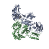
| ||||||||
|---|---|---|---|---|---|---|---|---|---|
| 1 |
| ||||||||
| Unit cell |
|
-Components
| #1: Protein | Mass: 63957.176 Da / Num. of mol.: 1 / Fragment: UNP residues 600-1154 / Mutation: K101P, K172A, K173A, C280S Source method: isolated from a genetically manipulated source Details: Lentivirus Source: (gene. exp.) Human immunodeficiency virus type 1 group M subtype B (isolate BH10)Strain: isolate BH10 / Gene: gag-pol / Plasmid: pCDF-2 / Production host:  References: UniProt: P03366, HIV-1 retropepsin, RNA-directed DNA polymerase, DNA-directed DNA polymerase, retroviral ribonuclease H, exoribonuclease H |
|---|---|
| #2: Protein | Mass: 50039.488 Da / Num. of mol.: 1 / Fragment: UNP residues 600-1027 / Mutation: C280S Source method: isolated from a genetically manipulated source Details: Lentivirus Source: (gene. exp.) Human immunodeficiency virus type 1 group M subtype B (isolate BH10)Strain: isolate BH10 / Gene: gag-pol / Plasmid: pCDF-2 / Production host: References: UniProt: P03366, HIV-1 retropepsin, RNA-directed DNA polymerase, DNA-directed DNA polymerase, retroviral ribonuclease H, exoribonuclease H |
| #3: Chemical | ChemComp-29T /   Mass: 388.376 Da / Num. of mol.: 1 / Source method: obtained synthetically / Formula: C21H16N4O4 Mass: 388.376 Da / Num. of mol.: 1 / Source method: obtained synthetically / Formula: C21H16N4O4 |
-Experimental details
-Experiment
| Experiment | Method: X-RAY DIFFRACTION / Number of used crystals: 1 |
|---|
- Sample preparation
| Crystal | Density Matthews: 3.39 Å3/Da / Density % sol: 63.75 % |
|---|---|
| Crystal grow | Temperature: 277 K / Method: vapor diffusion, hanging drop / pH: 7 Details: co-crystallization; 15% PEG 8000 (50% (w/v)), in pH 7.0 solution (0.125 M HEPES, 0.0125 M spermine-HCl, 0.04 M magnesium sulfate, 0.04 M ammonium sulfate) |
-Data collection
| Diffraction | Mean temperature: 80 K |
|---|---|
| Diffraction source | Source: SYNCHROTRON / Site: NSLS / Beamline: X29A / Wavelength: 1.075 Å |
| Detector | Type: ADSC QUANTUM 315r / Detector: CCD / Date: Jun 27, 2014 |
| Radiation | Monochromator: Si(111) / Protocol: SINGLE WAVELENGTH / Monochromatic (M) / Laue (L): M / Scattering type: x-ray |
| Radiation wavelength | Wavelength: 1.075 Å / Relative weight: 1 |
| Reflection | Resolution: 3.5→50 Å / Num. obs: 23862 / % possible obs: 100 % / Observed criterion σ(I): -3 / Redundancy: 3.7 % / Rsym value: 0.083 / Net I/σ(I): 24.1 |
| Reflection shell | Resolution: 3.5→3.56 Å / Redundancy: 3.7 % / Rmerge(I) obs: 0.603 / Mean I/σ(I) obs: 1.94 / % possible all: 100 |
- Processing
| Software |
| |||||||||||||||||||||||||||||||||||||||||||||||||||||||||||||||||||||||||||||||||||||||||||
|---|---|---|---|---|---|---|---|---|---|---|---|---|---|---|---|---|---|---|---|---|---|---|---|---|---|---|---|---|---|---|---|---|---|---|---|---|---|---|---|---|---|---|---|---|---|---|---|---|---|---|---|---|---|---|---|---|---|---|---|---|---|---|---|---|---|---|---|---|---|---|---|---|---|---|---|---|---|---|---|---|---|---|---|---|---|---|---|---|---|---|---|---|
| Refinement | Resolution: 3.5→38.223 Å / SU ML: 0.59 / Cross valid method: FREE R-VALUE / σ(F): 1.35 / Phase error: 33.94 / Stereochemistry target values: ML
| |||||||||||||||||||||||||||||||||||||||||||||||||||||||||||||||||||||||||||||||||||||||||||
| Solvent computation | Shrinkage radii: 0.9 Å / VDW probe radii: 1.11 Å / Solvent model: FLAT BULK SOLVENT MODEL | |||||||||||||||||||||||||||||||||||||||||||||||||||||||||||||||||||||||||||||||||||||||||||
| Refinement step | Cycle: LAST / Resolution: 3.5→38.223 Å
| |||||||||||||||||||||||||||||||||||||||||||||||||||||||||||||||||||||||||||||||||||||||||||
| Refine LS restraints |
| |||||||||||||||||||||||||||||||||||||||||||||||||||||||||||||||||||||||||||||||||||||||||||
| LS refinement shell |
|
