 Movie
Movie Controller

[English] 日本語
 Yorodumi
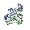
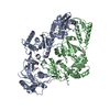
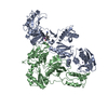
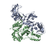
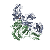
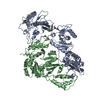
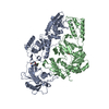
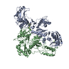
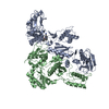
- PDB-2i5j: Crystal structure of HIV-1 reverse transcriptase (RT) in complex ... -
+ Open data

- Basic information
| Entry | Database: PDB / ID: 2i5j | |||||||||
|---|---|---|---|---|---|---|---|---|---|---|
| Title | Crystal structure of HIV-1 reverse transcriptase (RT) in complex with DHBNH, an RNASE H inhibitor | |||||||||
 Components Components | (Reverse transcriptase/ribonuclease H ...) x 2 | |||||||||
 Keywords Keywords | TRANSFERASE / AIDS / HIV / REVERSE TRANSCRIPTASE / RT / RNase H Inhibitor / RNHI / STRUCTURE-BASED DRUG DESIGN / PROTEIN-INHIBITOR COMPLEX / DRUG RESISTANCE | |||||||||
| Function / homology |  Function and homology information Function and homology informationHIV-1 retropepsin / symbiont-mediated activation of host apoptosis / retroviral ribonuclease H / exoribonuclease H / exoribonuclease H activity / DNA integration / viral genome integration into host DNA / establishment of integrated proviral latency / RNA-directed DNA polymerase / RNA stem-loop binding ...HIV-1 retropepsin / symbiont-mediated activation of host apoptosis / retroviral ribonuclease H / exoribonuclease H / exoribonuclease H activity / DNA integration / viral genome integration into host DNA / establishment of integrated proviral latency / RNA-directed DNA polymerase / RNA stem-loop binding / viral penetration into host nucleus / host multivesicular body / RNA-directed DNA polymerase activity / RNA-DNA hybrid ribonuclease activity / Transferases; Transferring phosphorus-containing groups; Nucleotidyltransferases / host cell / viral nucleocapsid / endonuclease activity / DNA recombination / DNA-directed DNA polymerase / aspartic-type endopeptidase activity / Hydrolases; Acting on ester bonds / DNA-directed DNA polymerase activity / symbiont-mediated suppression of host gene expression / viral translational frameshifting / symbiont entry into host cell / lipid binding / host cell nucleus / host cell plasma membrane / virion membrane / structural molecule activity / proteolysis / DNA binding / zinc ion binding Similarity search - Function | |||||||||
| Biological species |   Human immunodeficiency virus 1 Human immunodeficiency virus 1 | |||||||||
| Method |  X-RAY DIFFRACTION / SYNCHROTRON / MOLECULAR REPLACEMENT / Resolution: 3.15 Å X-RAY DIFFRACTION / SYNCHROTRON / MOLECULAR REPLACEMENT / Resolution: 3.15 Å | |||||||||
 Authors Authors | Himmel, D.M. / Sarafianos, S.G. / Knight, J.L. / Levy, R.M. / Arnold, E. | |||||||||
 Citation Citation | Journal: Acs Chem.Biol. / Year: 2006 Title: HIV-1 reverse transcriptase structure with RNase H inhibitor dihydroxy benzoyl naphthyl hydrazone bound at a novel site. Authors: Himmel, D.M. / Sarafianos, S.G. / Dharmasena, S. / Hossain, M.M. / McCoy-Simandle, K. / Ilina, T. / Clark, A.D. / Knight, J.L. / Julias, J.G. / Clark, P.K. / Krogh-Jespersen, K. / Levy, R.M. ...Authors: Himmel, D.M. / Sarafianos, S.G. / Dharmasena, S. / Hossain, M.M. / McCoy-Simandle, K. / Ilina, T. / Clark, A.D. / Knight, J.L. / Julias, J.G. / Clark, P.K. / Krogh-Jespersen, K. / Levy, R.M. / Hughes, S.H. / Parniak, M.A. / Arnold, E. #1: Journal: J.Med.Chem. / Year: 2005Title: Crystal Structures for HIV-1 Reverse Transcriptase in Complexes with Three Pyridinone Derivatives: A New Class of Non-nucleoside Inhibitors Effective Against a Broad Range of Drug-Resistant Strains Authors: Himmel, D.M. / Das, K. / Clark Jr., A.D. / Hughes, S.H. / Benjahad, A. / Oumouch, S. / Guillemont, J. / Coupa, S. / Poncelet, A. / Csoka, I. / Meyer, C. / Andries, K. / Nguyen, C.H. / Grierson, D.S. / Arnold, E. #2: Journal: EMBO J. / Year: 2001Title: Crystal structure of HIV-1 reverse transcriptase in complex with a polypurine tract RNA:DNA Authors: Sarafianos, S.G. / Das, K. / Tantillo, C. / Clark Jr., A.D. / Ding, J. / Whitcomb, J.M. / Boyer, P.L. / Hughes, S.H. / Arnold, E. #3: Journal: J.Med.Chem. / Year: 2004Title: Roles of Conformational and Positional Adaptability in Structure-Based Design of Tmc125-R165335 (Etravirine) and Related Non-Nucleoside Reverse Transcriptase Inhibitors that are Highly Potent ...Title: Roles of Conformational and Positional Adaptability in Structure-Based Design of Tmc125-R165335 (Etravirine) and Related Non-Nucleoside Reverse Transcriptase Inhibitors that are Highly Potent and Effective Against Wild-Type and Drug-Resistant HIV-1 Variants Authors: Das, K. / Clark Jr., A.D. / Lewi, P.J. / Heeres, J. / De Jonge, M.R. / Koymans, L.M.H. / Vinkers, H.M. / Daeyaert, F. / Ludovici, D.W. / Kukla, M.J. / De Corte, B. / Kavash, R.W. / Ho, C.Y. ...Authors: Das, K. / Clark Jr., A.D. / Lewi, P.J. / Heeres, J. / De Jonge, M.R. / Koymans, L.M.H. / Vinkers, H.M. / Daeyaert, F. / Ludovici, D.W. / Kukla, M.J. / De Corte, B. / Kavash, R.W. / Ho, C.Y. / Ye, H. / Lichtenstein, M.A. / Andries, K. / Pauwels, R. / De Bethune, M.-P. / Boyer, P.L. / Clark, P. / Hughes, S.H. / Janssen, P.A.J. / Arnold, E. #4: Journal: J.Mol.Biol. / Year: 1998Title: Structure and Functional Implications of the Polymerase Active Site Region in a Complex of HIV-1 RT with a Double-Stranded DNA Template-Primer and an Antibody Fab Fragment at 2.8 A Resolution. Authors: Ding, J. / Das, K. / Hsiou, Y. / Sarafianos, S.G. / Clark Jr., A.D. / Jacobo-Molina, A. / Tantillo, C. / Hughes, S.H. / Arnold, E. #5: Journal: Structure / Year: 1996Title: Structure of unliganded HIV-1 reverse transcriptase t 2.7 A resolution: implications of conformational hanges for polymerization and inhibition mechanisms Authors: Hsiou, Y. / Ding, J. / Das, K. / Clark Jr., A.D. / Hughes, S.H. / Arnold, E. | |||||||||
| History |
|
- Structure visualization
| Structure viewer | Molecule: MolmilJmol/JSmol |
|---|
- Downloads & links
-Download
| PDBx/mmCIF format | 2i5j.cif.gz | 217.5 KB | Display | PDBx/mmCIF format |
|---|---|---|---|---|
| PDB format | pdb2i5j.ent.gz | 170.5 KB | Display | PDB format |
| PDBx/mmJSON format | 2i5j.json.gz | Tree view | PDBx/mmJSON format | |
| Others |  Other downloads Other downloads |
-Validation report
| Arichive directory | https://data.pdbj.org/pub/pdb/validation_reports/i5/2i5jftp://data.pdbj.org/pub/pdb/validation_reports/i5/2i5j | HTTPS FTP |
|---|
-Related structure data
| Related structure data | 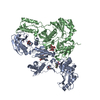 2be2S S: Starting model for refinement |
|---|---|
| Similar structure data |

-Links
 PDBj
PDBj


- Assembly
| Deposited unit | 
| ||||||||
|---|---|---|---|---|---|---|---|---|---|
| 1 |
| ||||||||
| Unit cell |
|
-Components
-Reverse transcriptase/ribonuclease H ... , 2 types, 2 molecules AB
| #1: Protein | Mass: 63659.906 Da / Num. of mol.: 1 / Fragment: residues 599-1150 / Mutation: C280S Source method: isolated from a genetically manipulated source Source: (gene. exp.) Human immunodeficiency virus 1 / Genus: Lentivirus / Strain: ISOLATE BH10 / Gene: gag / Production host:  |
|---|---|
| #2: Protein | Mass: 50152.648 Da / Num. of mol.: 1 / Fragment: residues 599-1027 / Mutation: C280S Source method: isolated from a genetically manipulated source Source: (gene. exp.) Human immunodeficiency virus 1 / Genus: Lentivirus / Strain: ISOLATE BH10 / Gene: gag / Production host: References: UniProt: P03366, UniProt: Q8UM83*PLUS, RNA-directed DNA polymerase |
-Sugars , 2 types, 5 molecules 
| #3: Polysaccharide |   Source method: isolated from a genetically manipulated source Details: oligosaccharide with reducing-end-to-reducing-end glycosidic bond References: sucrose #4: Sugar |  Type: D-saccharide, alpha linking / Mass: 180.156 Da / Num. of mol.: 3 Type: D-saccharide, alpha linking / Mass: 180.156 Da / Num. of mol.: 3Source method: isolated from a genetically manipulated source Formula: C6H12O6 |
|---|
-Non-polymers , 3 types, 38 molecules 


| #5: Chemical | ChemComp-K05 / ( Mass: 336.341 Da / Num. of mol.: 1 / Source method: obtained synthetically / Formula: C19H16N2O4 Mass: 336.341 Da / Num. of mol.: 1 / Source method: obtained synthetically / Formula: C19H16N2O4 |
|---|---|
| #6: Chemical | ChemComp-MG /  Mass: 24.305 Da / Num. of mol.: 1 / Source method: obtained synthetically / Formula: Mg Mass: 24.305 Da / Num. of mol.: 1 / Source method: obtained synthetically / Formula: Mg |
| #7: Water | ChemComp-HOH / Mass: 18.015 Da / Num. of mol.: 36 / Source method: isolated from a natural source / Formula: H2O |
-Experimental details
-Experiment
| Experiment | Method: X-RAY DIFFRACTION / Number of used crystals: 1 |
|---|
- Sample preparation
| Crystal | Density Matthews: 3.41 Å3/Da / Density % sol: 63.95 % |
|---|---|
| Crystal grow | Temperature: 277 K / Method: vapor diffusion, hanging drop / pH: 6.4 Details: 50 mM Imidazole pH 6.4, 100 mM Ammonium Sulfate, 15 mM Magnesium Sulfate, 0.6 mM DHBNH, 4% Glucose, 10.5% PEG 8000, 2 mM NaN3, VAPOR DIFFUSION, HANGING DROP, temperature 277K |
-Data collection
| Diffraction | Mean temperature: 100 K |
|---|---|
| Diffraction source | Source: SYNCHROTRON / Site: NSLS  / Beamline: X25 / Wavelength: 1.1 Å / Beamline: X25 / Wavelength: 1.1 Å |
| Detector | Type: ADSC QUANTUM 315 / Detector: CCD / Date: Jun 4, 2003 |
| Radiation | Protocol: SINGLE WAVELENGTH / Monochromatic (M) / Laue (L): M / Scattering type: x-ray |
| Radiation wavelength | Wavelength: 1.1 Å / Relative weight: 1 |
| Reflection | Resolution: 3.15→26 Å / Num. all: 27482 / Num. obs: 24789 / % possible obs: 90.2 % / Observed criterion σ(F): 0 / Observed criterion σ(I): -1 / Redundancy: 5.2 % / Biso Wilson estimate: 66.849 Å2 / Rsym value: 0.101 / Χ2: 1.001 / Net I/σ(I): 11.1 |
| Reflection shell | Resolution: 3.15→3.26 Å / Redundancy: 2.8 % / Num. unique all: 2011 / Rsym value: 0.805 / Χ2: 0.847 / % possible all: 74.4 |
-Phasing
| Phasing MR | Rfactor: 0.365 / R rigid body: 0.351 / Cor.coef. Fo:Fc: 0.759 / Cor.coef. Io to Ic: 0.683
| ||||||
|---|---|---|---|---|---|---|---|
| Phasing dm shell | Resolution: 3→25 Å / Delta phi final: 0.123 / FOM : 0.125 / Reflection: 25136 |
- Processing
| Software |
| ||||||||||||||||||||||||||||||||||||
|---|---|---|---|---|---|---|---|---|---|---|---|---|---|---|---|---|---|---|---|---|---|---|---|---|---|---|---|---|---|---|---|---|---|---|---|---|---|
| Refinement | Method to determine structure: MOLECULAR REPLACEMENT Starting model: PDB ENTRY 2BE2 Resolution: 3.15→25.71 Å / Rfactor Rfree error: 0.01 / FOM work R set: 0.819 / Data cutoff high absF: 197616.641 / Data cutoff low absF: 0 / Isotropic thermal model: RESTRAINED / Cross valid method: THROUGHOUT / σ(F): 0 / Stereochemistry target values: Engh & Huber
| ||||||||||||||||||||||||||||||||||||
| Solvent computation | Solvent model: FLAT MODEL / Bsol: 52.394 Å2 / ksol: 0.277 e/Å3 | ||||||||||||||||||||||||||||||||||||
| Displacement parameters | Biso mean: 92.2 Å2
| ||||||||||||||||||||||||||||||||||||
| Refine analyze |
| ||||||||||||||||||||||||||||||||||||
| Refinement step | Cycle: LAST / Resolution: 3.15→25.71 Å
| ||||||||||||||||||||||||||||||||||||
| Refine LS restraints |
| ||||||||||||||||||||||||||||||||||||
| LS refinement shell | Resolution: 3.15→3.35 Å / Rfactor Rfree error: 0.049 / Total num. of bins used: 6
| ||||||||||||||||||||||||||||||||||||
| Xplor file |
|
