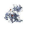
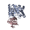
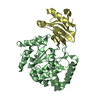
- PDB-5ays: Crystal structure of SAUGI/HSV UDG complex -
+
Open data
ID or keywords:
Loading...
-
Basic information
Entry
Database: PDB / ID: 5ays
Title
Crystal structure of SAUGI/HSV UDG complex
Components
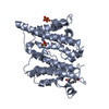
Uncharacterized protein
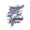
Uracil-DNA glycosylase
Keywords
HYDROLASE INHIBITOR / DNA mimic protein / DNA mimicking / uracil-DNA glycosylase inhibitor / uracil-DNA glycosylase / Herpes simplex virus
Function / homology
Function and homology information
base-excision repair, AP site formation via deaminated base removal / uracil-DNA glycosylase / uracil DNA N-glycosylase activity / symbiont-mediated perturbation of host defense response / host cell nucleus / metal ion binding Similarity search - Function
S. aureus uracil DNA glycosylase inhibitor / S. aureus uracil DNA glycosylase inhibitor / Uracil DNA glycosylase inhibitor superfamily / S. aureus uracil DNA glycosylase inhibitor / Uracil-DNA glycosylase family 1 / Uracil DNA glycosylase superfamily / UreE urease accessory protein, C-terminal domain / Uracil-DNA Glycosylase, subunit E / Uracil-DNA glycosylase-like domain / Uracil-DNA glycosylase, active site ...S. aureus uracil DNA glycosylase inhibitor / S. aureus uracil DNA glycosylase inhibitor / Uracil DNA glycosylase inhibitor superfamily / S. aureus uracil DNA glycosylase inhibitor / Uracil-DNA glycosylase family 1 / Uracil DNA glycosylase superfamily / UreE urease accessory protein, C-terminal domain / Uracil-DNA Glycosylase, subunit E / Uracil-DNA glycosylase-like domain / Uracil-DNA glycosylase, active site / Uracil-DNA glycosylase signature. / Uracil-DNA glycosylase-like / Uracil DNA glycosylase superfamily / Uracil-DNA glycosylase-like domain superfamily / Nuclear Transport Factor 2; Chain: A, / Roll / 3-Layer(aba) Sandwich / Alpha Beta Similarity search - Domain/homology
Resolution: 2.09→20 Å / Cor.coef. Fo:Fc: 0.948 / Cor.coef. Fo:Fc free: 0.933 / SU B: 4.807 / SU ML: 0.128 / Cross valid method: THROUGHOUT / ESU R: 0.261 / ESU R Free: 0.187 / Stereochemistry target values: MAXIMUM LIKELIHOOD / Details: HYDROGENS HAVE BEEN ADDED IN THE RIDING POSITIONS
Rfactor
Num. reflection
% reflection
Selection details
Rfree
0.22253
2191
5 %
RANDOM
Rwork
0.19486
-
-
-
obs
0.19629
41236
94.38 %
-
Solvent computation
Ion probe radii: 0.8 Å / Shrinkage radii: 0.8 Å / VDW probe radii: 1.4 Å / Solvent model: MASK
 Movie
Movie Controller


 Open data
 Basic information
 Components
 Keywords
 Function and homology information
 Human herpesvirus 1 (Herpes simplex virus type 1)
 Staphylococcus aureus (bacteria)
 X-RAY DIFFRACTION /
  Authors
 Taiwan, 2items
  Citation
 Structure visualization
 Downloads & links
 Other downloads
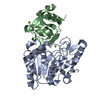


 PDBj
PDBj Assembly
 Mass: 18.015 Da / Num. of mol.: 712 / Source method: isolated from a natural source / Formula: H2O
 Sample preparation
 Processing