 Movie
Movie Controller


+ Open data

- Basic information
| Entry | Database: PDB / ID: 1lau | ||||||
|---|---|---|---|---|---|---|---|
| Title | URACIL-DNA GLYCOSYLASE | ||||||
 Components Components |
| ||||||
 Keywords Keywords | HYDROLASE/DNA / HYDROLASE / GLYCOSIDASE / DNA / HYDROLASE-DNA COMPLEX | ||||||
| Function / homology |  Function and homology information Function and homology informationbase-excision repair, AP site formation via deaminated base removal / uracil-DNA glycosylase / uracil DNA N-glycosylase activity / symbiont-mediated perturbation of host defense response / host cell nucleus Similarity search - Function | ||||||
| Biological species |   Human herpesvirus 1 (Herpes simplex virus type 1) Human herpesvirus 1 (Herpes simplex virus type 1) | ||||||
| Method |  X-RAY DIFFRACTION / Resolution: 1.8 Å X-RAY DIFFRACTION / Resolution: 1.8 Å | ||||||
 Authors Authors | Pearl, L.H. / Savva, R. | ||||||
 Citation Citation | Journal: Nature / Year: 1995 Title: The structural basis of specific base-excision repair by uracil-DNA glycosylase. Authors: Savva, R. / McAuley-Hecht, K. / Brown, T. / Pearl, L. #1: Journal: J.Mol.Biol. / Year: 1993Title: Crystallization and Preliminary X-Ray Analysis of the Uracil-DNA Glycosylase DNA Repair Enzyme from Herpes Simplex Virus Type 1 Authors: Savva, R. / Pearl, L.H. | ||||||
| History |
|
- Structure visualization
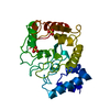
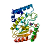
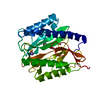
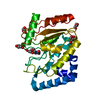
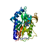
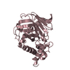
| Structure viewer | Molecule: MolmilJmol/JSmol |
|---|
- Downloads & links
-Download
| PDBx/mmCIF format | 1lau.cif.gz | 62.8 KB | Display | PDBx/mmCIF format |
|---|---|---|---|---|
| PDB format | pdb1lau.ent.gz | 45.7 KB | Display | PDB format |
| PDBx/mmJSON format | 1lau.json.gz | Tree view | PDBx/mmJSON format | |
| Others |  Other downloads Other downloads |
-Validation report
| Arichive directory | https://data.pdbj.org/pub/pdb/validation_reports/la/1lauftp://data.pdbj.org/pub/pdb/validation_reports/la/1lau | HTTPS FTP |
|---|
-Related structure data


-Links
 PDBj
PDBj

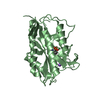
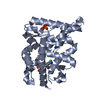
- Assembly
| Deposited unit | 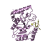
| ||||||||
|---|---|---|---|---|---|---|---|---|---|
| 1 |
| ||||||||
| Unit cell |
|
-Components
| #1: DNA chain | Mass: 867.621 Da / Num. of mol.: 1 / Source method: obtained synthetically |
|---|---|
| #2: Protein | Mass: 27366.447 Da / Num. of mol.: 1 / Source method: isolated from a natural source Source: (natural) Human herpesvirus 1 (Herpes simplex virus type 1)Genus: Simplexvirus / Strain: STRAIN 17 / References: UniProt: P10186 |
| #3: Water | ChemComp-HOH /  Mass: 18.015 Da / Num. of mol.: 161 / Source method: isolated from a natural source / Formula: H2O Mass: 18.015 Da / Num. of mol.: 161 / Source method: isolated from a natural source / Formula: H2O |
| Compound details | THE URACIL-DNA GLYCOSYLASE MOLECULE IN THIS STUDY CONSISTS OF 244 AMINO ACIDS FROM THE METHIONINE ...THE URACIL-DNA GLYCOSYLAS |
-Experimental details
-Experiment
| Experiment | Method: X-RAY DIFFRACTION |
|---|
- Sample preparation
| Crystal | Density Matthews: 2.21 Å3/Da / Density % sol: 44.4 % | |||||||||||||||||||||||||
|---|---|---|---|---|---|---|---|---|---|---|---|---|---|---|---|---|---|---|---|---|---|---|---|---|---|---|
| Crystal grow | *PLUS pH: 6.8 / Method: unknown | |||||||||||||||||||||||||
| Components of the solutions | *PLUS
|
-Data collection
| Detector | Type: MARRESEARCH / Detector: IMAGE PLATE |
|---|---|
| Radiation | Protocol: SINGLE WAVELENGTH / Monochromatic (M) / Laue (L): M / Scattering type: x-ray |
| Radiation wavelength | Relative weight: 1 |
| Reflection | Num. obs: 20152 / % possible obs: 90.3 % / Redundancy: 2.1 % |
| Reflection | *PLUS % possible obs: 90.3 % / Redundancy: 2.1 % |
- Processing
| Software |
| ||||||||||||||||||||||||||||||||||||||||||||||||||||||||||||
|---|---|---|---|---|---|---|---|---|---|---|---|---|---|---|---|---|---|---|---|---|---|---|---|---|---|---|---|---|---|---|---|---|---|---|---|---|---|---|---|---|---|---|---|---|---|---|---|---|---|---|---|---|---|---|---|---|---|---|---|---|---|
| Refinement | Resolution: 1.8→8 Å / σ(F): 0
| ||||||||||||||||||||||||||||||||||||||||||||||||||||||||||||
| Refinement step | Cycle: LAST / Resolution: 1.8→8 Å
| ||||||||||||||||||||||||||||||||||||||||||||||||||||||||||||
| Refine LS restraints |
| ||||||||||||||||||||||||||||||||||||||||||||||||||||||||||||
| Software | *PLUS Name: X-PLOR / Classification: refinement | ||||||||||||||||||||||||||||||||||||||||||||||||||||||||||||
| Refinement | *PLUS Highest resolution: 1.8 Å / Lowest resolution: 8 Å / σ(F): 0 | ||||||||||||||||||||||||||||||||||||||||||||||||||||||||||||
| Solvent computation | *PLUS | ||||||||||||||||||||||||||||||||||||||||||||||||||||||||||||
| Displacement parameters | *PLUS |
