 Movie
Movie Controller

[English] 日本語
 Yorodumi
- PDB-5aam: Structure of a redesigned cross-reactive antibody to dengue virus... -
+ Open data

- Basic information
| Entry | Database: PDB / ID: 5aam | |||||||||
|---|---|---|---|---|---|---|---|---|---|---|
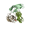
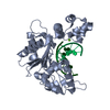
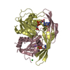
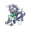
| Title | Structure of a redesigned cross-reactive antibody to dengue virus with increased in vivo potency | |||||||||
 Components Components |
| |||||||||
 Keywords Keywords | IMMUNE SYSTEM / SCFV DENGUE ANTIBODY ENVELOPE DOMAIN III | |||||||||
| Function / homology |  Function and homology information Function and homology information: / viral capsid / clathrin-dependent endocytosis of virus by host cell / protein dimerization activity / symbiont-mediated suppression of host innate immune response / host cell endoplasmic reticulum membrane / fusion of virus membrane with host endosome membrane / viral envelope / virion attachment to host cell / host cell nucleus ...: / viral capsid / clathrin-dependent endocytosis of virus by host cell / protein dimerization activity / symbiont-mediated suppression of host innate immune response / host cell endoplasmic reticulum membrane / fusion of virus membrane with host endosome membrane / viral envelope / virion attachment to host cell / host cell nucleus / virion membrane / structural molecule activity / extracellular region / membrane Similarity search - Function | |||||||||
| Biological species |    DENGUE VIRUS DENGUE VIRUS | |||||||||
| Method |  X-RAY DIFFRACTION / SYNCHROTRON / MOLECULAR REPLACEMENT / Resolution: 2.49 Å X-RAY DIFFRACTION / SYNCHROTRON / MOLECULAR REPLACEMENT / Resolution: 2.49 Å | |||||||||
 Authors Authors | Wong, Y.H. / Robinson, L.N. / Lescar, J. / Sasisekharan, R. | |||||||||
 Citation Citation | Journal: Cell(Cambridge,Mass.) / Year: 2015 Title: Structure-Guided Design of an Anti-Dengue Antibody Directed to a Non-Immunodominant Epitope. Authors: Robinson, L.N. / Tharakaraman, K. / Rowley, K.J. / Costa, V.V. / Chan, K.R. / Wong, Y.H. / Ong, L.C. / Tan, H.C. / Koch, T. / Cain, D. / Kirloskar, R. / Viswanathan, K. / Liew, C.W. / ...Authors: Robinson, L.N. / Tharakaraman, K. / Rowley, K.J. / Costa, V.V. / Chan, K.R. / Wong, Y.H. / Ong, L.C. / Tan, H.C. / Koch, T. / Cain, D. / Kirloskar, R. / Viswanathan, K. / Liew, C.W. / Tissire, H. / Ramakrishnan, B. / Myette, J.R. / Babcock, G.J. / Sasisekharan, V. / Alonso, S. / Chen, J. / Lescar, J. / Shriver, Z. / Ooi, E.E. / Sasisekharan, R. | |||||||||
| History |
|
- Structure visualization
| Structure viewer | Molecule: MolmilJmol/JSmol |
|---|
- Downloads & links
-Download
| PDBx/mmCIF format | 5aam.cif.gz | 140.1 KB | Display | PDBx/mmCIF format |
|---|---|---|---|---|
| PDB format | pdb5aam.ent.gz | 109.6 KB | Display | PDB format |
| PDBx/mmJSON format | 5aam.json.gz | Tree view | PDBx/mmJSON format | |
| Others |  Other downloads Other downloads |
-Validation report
| Arichive directory | https://data.pdbj.org/pub/pdb/validation_reports/aa/5aamftp://data.pdbj.org/pub/pdb/validation_reports/aa/5aam | HTTPS FTP |
|---|
-Related structure data
| Related structure data |  5aawC  3uypS S: Starting model for refinement C: citing same article ( |
|---|---|
| Similar structure data |

-Links
 PDBj
PDBj


- Assembly
| Deposited unit | 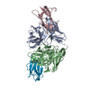
| ||||||||
|---|---|---|---|---|---|---|---|---|---|
| 1 | 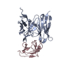
| ||||||||
| 2 | 
| ||||||||
| Unit cell |
|
-Components
| #1: Antibody | Mass: 27299.357 Da / Num. of mol.: 2 / Fragment: SCFV, RESIDUES 2-253 Source method: isolated from a genetically manipulated source Source: (gene. exp.)  #2: Protein | Mass: 12692.560 Da / Num. of mol.: 2 / Fragment: RESIDUES 2-109 Source method: isolated from a genetically manipulated source Source: (gene. exp.) DENGUE VIRUS / Production host: #3: Water | ChemComp-HOH / |  Mass: 18.015 Da / Num. of mol.: 127 / Source method: isolated from a natural source / Formula: H2O Mass: 18.015 Da / Num. of mol.: 127 / Source method: isolated from a natural source / Formula: H2OHas protein modification | Y | |
|---|
-Experimental details
-Experiment
| Experiment | Method: X-RAY DIFFRACTION / Number of used crystals: 1 |
|---|
- Sample preparation
| Crystal | Density Matthews: 2.11 Å3/Da / Density % sol: 41.67 % / Description: NONE |
|---|
-Data collection
| Diffraction | Mean temperature: 100 K |
|---|---|
| Diffraction source | Source: SYNCHROTRON / Site: SLS  / Beamline: X10SA / Wavelength: 1 / Beamline: X10SA / Wavelength: 1 |
| Detector | Type: DECTRIS PILATUS / Detector: PIXEL / Date: Nov 29, 2014 / Details: MIRRORS |
| Radiation | Protocol: SINGLE WAVELENGTH / Monochromatic (M) / Laue (L): M / Scattering type: x-ray |
| Radiation wavelength | Wavelength: 1 Å / Relative weight: 1 |
| Reflection | Resolution: 2.49→47.9 Å / Num. obs: 23183 / % possible obs: 98.3 % / Observed criterion σ(I): 0 / Redundancy: 6.8 % / Biso Wilson estimate: 80.51 Å2 / Rmerge(I) obs: 0.08 / Net I/σ(I): 14.8 |
| Reflection shell | Resolution: 2.49→2.63 Å / Redundancy: 6.7 % / Rmerge(I) obs: 1.15 / Mean I/σ(I) obs: 1.4 / % possible all: 92.3 |
- Processing
| Software |
| ||||||||||||||||||||||||||||||||||||||||||||||||||||||||||||||||||||||||||||||||||||||||||||||||||||||||||||||||||
|---|---|---|---|---|---|---|---|---|---|---|---|---|---|---|---|---|---|---|---|---|---|---|---|---|---|---|---|---|---|---|---|---|---|---|---|---|---|---|---|---|---|---|---|---|---|---|---|---|---|---|---|---|---|---|---|---|---|---|---|---|---|---|---|---|---|---|---|---|---|---|---|---|---|---|---|---|---|---|---|---|---|---|---|---|---|---|---|---|---|---|---|---|---|---|---|---|---|---|---|---|---|---|---|---|---|---|---|---|---|---|---|---|---|---|---|
| Refinement | Method to determine structure: MOLECULAR REPLACEMENT Starting model: PDB ENTRY 3UYP Resolution: 2.49→29.55 Å / Cor.coef. Fo:Fc: 0.9426 / Cor.coef. Fo:Fc free: 0.9169 / SU R Cruickshank DPI: 0.766 / Cross valid method: THROUGHOUT / σ(F): 0 / SU R Blow DPI: 0.738 / SU Rfree Blow DPI: 0.311 / SU Rfree Cruickshank DPI: 0.317
| ||||||||||||||||||||||||||||||||||||||||||||||||||||||||||||||||||||||||||||||||||||||||||||||||||||||||||||||||||
| Displacement parameters | Biso mean: 85.63 Å2
| ||||||||||||||||||||||||||||||||||||||||||||||||||||||||||||||||||||||||||||||||||||||||||||||||||||||||||||||||||
| Refine analyze | Luzzati coordinate error obs: 0.428 Å | ||||||||||||||||||||||||||||||||||||||||||||||||||||||||||||||||||||||||||||||||||||||||||||||||||||||||||||||||||
| Refinement step | Cycle: LAST / Resolution: 2.49→29.55 Å
| ||||||||||||||||||||||||||||||||||||||||||||||||||||||||||||||||||||||||||||||||||||||||||||||||||||||||||||||||||
| Refine LS restraints |
| ||||||||||||||||||||||||||||||||||||||||||||||||||||||||||||||||||||||||||||||||||||||||||||||||||||||||||||||||||
| LS refinement shell | Resolution: 2.49→2.6 Å / Total num. of bins used: 12
|
