 Movie
Movie Controller


+ Open data

- Basic information
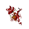
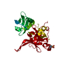
| Entry | Database: PDB / ID: 5to0 | ||||||
|---|---|---|---|---|---|---|---|
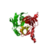
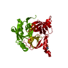
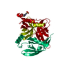
| Title | HTRA2 S276C mutant | ||||||
 Components Components | Serine protease HTRA2, mitochondrial | ||||||
 Keywords Keywords | HYDROLASE / Serine Protease / Parkinson Disease / Mitochondria / PDZ Dynamics | ||||||
| Function / homology |  Function and homology information Function and homology informationHtrA2 peptidase / pentacyclic triterpenoid metabolic process / negative regulation of type 2 mitophagy / regulation of autophagy of mitochondrion / ceramide metabolic process / CD40 receptor complex / mitochondrial protein catabolic process / programmed cell death / positive regulation of extrinsic apoptotic signaling pathway in absence of ligand / serine-type endopeptidase complex ...HtrA2 peptidase / pentacyclic triterpenoid metabolic process / negative regulation of type 2 mitophagy / regulation of autophagy of mitochondrion / ceramide metabolic process / CD40 receptor complex / mitochondrial protein catabolic process / programmed cell death / positive regulation of extrinsic apoptotic signaling pathway in absence of ligand / serine-type endopeptidase complex / adult walking behavior / response to herbicide / execution phase of apoptosis / : / forebrain development / protein autoprocessing / negative regulation of cell cycle / regulation of multicellular organism growth / Mitochondrial unfolded protein response (UPRmt) / positive regulation of execution phase of apoptosis / protein serine/threonine kinase inhibitor activity / ubiquitin ligase inhibitor activity / negative regulation of oxidative stress-induced intrinsic apoptotic signaling pathway / neuron development / cellular response to retinoic acid / cellular response to interferon-beta / Mitochondrial protein degradation / serine-type peptidase activity / protein catabolic process / mitochondrion organization / mitochondrial intermembrane space / cellular response to growth factor stimulus / mitochondrial membrane / intrinsic apoptotic signaling pathway in response to DNA damage / cytoplasmic side of plasma membrane / : / peptidase activity / cellular response to heat / cellular response to oxidative stress / neuron apoptotic process / cytoskeleton / negative regulation of neuron apoptotic process / intracellular signal transduction / positive regulation of apoptotic process / serine-type endopeptidase activity / endoplasmic reticulum membrane / chromatin / endoplasmic reticulum / mitochondrion / proteolysis / membrane / identical protein binding / nucleus / cytosol Similarity search - Function | ||||||
| Biological species |  Homo sapiens (human) Homo sapiens (human) | ||||||
| Method |  X-RAY DIFFRACTION / SYNCHROTRON / MOLECULAR REPLACEMENT / Resolution: 1.9 Å X-RAY DIFFRACTION / SYNCHROTRON / MOLECULAR REPLACEMENT / Resolution: 1.9 Å | ||||||
 Authors Authors | Merski, M. / Barbosa Pereira, P.J. / Macedo-Ribeiro, S. | ||||||
| Funding support |  Portugal, 1items Portugal, 1items
| ||||||
 Citation Citation | Journal: Cell Death Dis / Year: 2017 Title: Molecular motion regulates the activity of the Mitochondrial Serine Protease HtrA2. Authors: Merski, M. / Moreira, C. / Abreu, R.M. / Ramos, M.J. / Fernandes, P.A. / Martins, L.M. / Pereira, P.J.B. / Macedo-Ribeiro, S. | ||||||
| History |
|
- Structure visualization
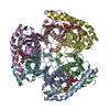
| Structure viewer | Molecule: MolmilJmol/JSmol |
|---|
- Downloads & links
-Download
| PDBx/mmCIF format | 5to0.cif.gz | 71.6 KB | Display | PDBx/mmCIF format |
|---|---|---|---|---|
| PDB format | pdb5to0.ent.gz | 51 KB | Display | PDB format |
| PDBx/mmJSON format | 5to0.json.gz | Tree view | PDBx/mmJSON format | |
| Others |  Other downloads Other downloads |
-Validation report
| Arichive directory | https://data.pdbj.org/pub/pdb/validation_reports/to/5to0ftp://data.pdbj.org/pub/pdb/validation_reports/to/5to0 | HTTPS FTP |
|---|
-Related structure data
| Related structure data |  5m3nC 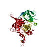 5m3oC  5tnyC  5tnzC  5to1C  1lcyS C: citing same article ( S: Starting model for refinement |
|---|---|
| Similar structure data |

-Links
 PDBj
PDBj- Assembly
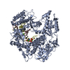
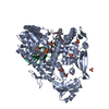
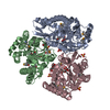
| Deposited unit | 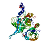
| ||||||||
|---|---|---|---|---|---|---|---|---|---|
| 1 | 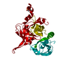
| ||||||||
| Unit cell |
|
-Components
| #1: Protein | Mass: 35993.949 Da / Num. of mol.: 1 / Mutation: Mutation S276C Source method: isolated from a genetically manipulated source Source: (gene. exp.) Homo sapiens (human) / Gene: HTRA2, OMI, PRSS25 / Production host:  |
|---|---|
| #2: Chemical | ChemComp-MES /   Mass: 195.237 Da / Num. of mol.: 1 / Source method: obtained synthetically / Formula: C6H13NO4S / Comment: pH buffer*YM Mass: 195.237 Da / Num. of mol.: 1 / Source method: obtained synthetically / Formula: C6H13NO4S / Comment: pH buffer*YM |
| #3: Water | ChemComp-HOH /  Mass: 18.015 Da / Num. of mol.: 75 / Source method: isolated from a natural source / Formula: H2O Mass: 18.015 Da / Num. of mol.: 75 / Source method: isolated from a natural source / Formula: H2O |
-Experimental details
-Experiment
| Experiment | Method: X-RAY DIFFRACTION / Number of used crystals: 1 |
|---|
- Sample preparation
| Crystal | Density Matthews: 2.38 Å3/Da / Density % sol: 48.28 % |
|---|---|
| Crystal grow | Temperature: 293 K / Method: vapor diffusion, sitting drop / pH: 6 Details: 0.1 M MES pH 6.0, 1 M LiCl, and 15-20% (w/v) PEG-6000 |
-Data collection
| Diffraction | Mean temperature: 100 K |
|---|---|
| Diffraction source | Source: SYNCHROTRON / Site: ESRF  / Beamline: ID14-3 / Wavelength: 0.931 Å / Beamline: ID14-3 / Wavelength: 0.931 Å |
| Detector | Type: ADSC QUANTUM 4 / Detector: CCD / Date: Jun 30, 2007 |
| Radiation | Protocol: SINGLE WAVELENGTH / Monochromatic (M) / Laue (L): M / Scattering type: x-ray |
| Radiation wavelength | Wavelength: 0.931 Å / Relative weight: 1 |
| Reflection | Resolution: 1.9→63.1 Å / Num. obs: 26268 / % possible obs: 99.8 % / Redundancy: 8.4 % / Rmerge(I) obs: 0.044 / Net I/σ(I): 30 |
| Reflection shell | Resolution: 1.9→2 Å / Redundancy: 8.4 % / Rmerge(I) obs: 0.512 / Mean I/σ(I) obs: 4.4 / Num. unique all: 3828 |
- Processing
| Software |
| |||||||||||||||||||||||||||||||||||||||||||||||||||||||||||||||||||||||||||||
|---|---|---|---|---|---|---|---|---|---|---|---|---|---|---|---|---|---|---|---|---|---|---|---|---|---|---|---|---|---|---|---|---|---|---|---|---|---|---|---|---|---|---|---|---|---|---|---|---|---|---|---|---|---|---|---|---|---|---|---|---|---|---|---|---|---|---|---|---|---|---|---|---|---|---|---|---|---|---|
| Refinement | Method to determine structure: MOLECULAR REPLACEMENT Starting model: 1LCY Resolution: 1.9→47.894 Å / Cross valid method: FREE R-VALUE / σ(F): 1.99 / Phase error: 30.57
| |||||||||||||||||||||||||||||||||||||||||||||||||||||||||||||||||||||||||||||
| Solvent computation | Shrinkage radii: 0.9 Å / VDW probe radii: 1.11 Å | |||||||||||||||||||||||||||||||||||||||||||||||||||||||||||||||||||||||||||||
| Refinement step | Cycle: LAST / Resolution: 1.9→47.894 Å
| |||||||||||||||||||||||||||||||||||||||||||||||||||||||||||||||||||||||||||||
| Refine LS restraints |
| |||||||||||||||||||||||||||||||||||||||||||||||||||||||||||||||||||||||||||||
| LS refinement shell |
|
