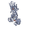
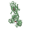
[English] 日本語
Yorodumi
- PDB-5a3s: Crystal structure of the (SR) Calcium ATPase E2-vanadate complex ... -

+
Open data


ID or keywords:

Loading...

-
Basic information

Entry
Database: PDB / ID: 5a3s
TitleCrystal structure of the (SR) Calcium ATPase E2-vanadate complex bound to thapsigargin and TNP-ATP
ComponentsSARCOPLASMIC RETICULUM CALCIUM ATPASE 1 MOLECULE SARCOPLASMIC/ENDOPLASMIC RETICULUM CALCIUM ATPASE 1
KeywordsHYDROLASE / SARCO(ENDO)PLASMIC RETICULUM CALCIUM ATPASE / BERYLLIUM FLUORIDE / P-TYPE ATPASE / SERCA / TRINITROPHENYL-NUCLEOTIDE ANALOGES / TNP-ATP / CALCIUM TRANSPORT / INHIBITION / TRANSITION STATE
Function / homology
Function and homology information


positive regulation of cardiac muscle cell contraction / positive regulation of calcium ion import into sarcoplasmic reticulum / H zone / positive regulation of fast-twitch skeletal muscle fiber contraction / calcium ion import into sarcoplasmic reticulum / negative regulation of striated muscle contraction / regulation of striated muscle contraction / positive regulation of ATPase-coupled calcium transmembrane transporter activity / P-type Ca2+ transporter / P-type calcium transporter activity ...positive regulation of cardiac muscle cell contraction / positive regulation of calcium ion import into sarcoplasmic reticulum / H zone / positive regulation of fast-twitch skeletal muscle fiber contraction / calcium ion import into sarcoplasmic reticulum / negative regulation of striated muscle contraction / regulation of striated muscle contraction / positive regulation of ATPase-coupled calcium transmembrane transporter activity / P-type Ca2+ transporter / P-type calcium transporter activity / I band / endoplasmic reticulum-Golgi intermediate compartment / sarcoplasmic reticulum membrane / sarcoplasmic reticulum / intracellular calcium ion homeostasis / calcium ion transport / calcium ion binding / endoplasmic reticulum membrane / perinuclear region of cytoplasm / endoplasmic reticulum / ATP hydrolysis activity / ATP binding / membrane
Similarity search - Function
Calcium-transporting ATPase, transmembrane domain / Calcium-transporting ATPase, transmembrane domain / P-type ATPase, subfamily IIA, SERCA-type / Calcium-transporting ATPase, cytoplasmic transduction domain A / Calcium-transporting ATPase, cytoplasmic transduction domain A / Calcium-transporting ATPase, cytoplasmic domain N / Calcium-transporting ATPase, cytoplasmic domain N / haloacid dehalogenase-like hydrolase / Cation-transporting P-type ATPase, C-terminal / Cation transporting ATPase, C-terminus ...Calcium-transporting ATPase, transmembrane domain / Calcium-transporting ATPase, transmembrane domain / P-type ATPase, subfamily IIA, SERCA-type / Calcium-transporting ATPase, cytoplasmic transduction domain A / Calcium-transporting ATPase, cytoplasmic transduction domain A / Calcium-transporting ATPase, cytoplasmic domain N / Calcium-transporting ATPase, cytoplasmic domain N / haloacid dehalogenase-like hydrolase / Cation-transporting P-type ATPase, C-terminal / Cation transporting ATPase, C-terminus / Cation transporter/ATPase, N-terminus / Cation-transporting P-type ATPase, N-terminal / Cation transporter/ATPase, N-terminus / Cation transport ATPase (P-type) / E1-E2 ATPase / P-type ATPase, haloacid dehalogenase domain / P-type ATPase, phosphorylation site / P-type ATPase, cytoplasmic domain N / E1-E2 ATPases phosphorylation site. / P-type ATPase, A domain superfamily / P-type ATPase / P-type ATPase, transmembrane domain superfamily / HAD superfamily/HAD-like / haloacid dehalogenase-like hydrolase / HAD superfamily / HAD-like superfamily / Distorted Sandwich / Up-down Bundle / Rossmann fold / 3-Layer(aba) Sandwich / Mainly Beta / Mainly Alpha / Alpha Beta
Similarity search - Domain/homology
Chem-128 / : / Chem-TG1 / oxido(dioxo)vanadium / Sarcoplasmic/endoplasmic reticulum calcium ATPase 1
Similarity search - Component
Biological speciesORYCTOLAGUS CUNICULUS (rabbit)
MethodX-RAY DIFFRACTION / SYNCHROTRON / MOLECULAR REPLACEMENT / Resolution: 3.3 Å
AuthorsClausen, J.D. / Bublitz, M. / Arnou, B. / Olesen, C. / Andersen, J.P. / Moller, J.V. / Nissen, P.
CitationJournal: Structure / Year: 2016
Title: Crystal Structure of the Vanadate-Inhibited Ca(2+)-ATPase.
Authors: Clausen, J.D. / Bublitz, M. / Arnou, B. / Olesen, C. / Andersen, J.P. / Moller, J.V. / Nissen, P.
History
DepositionJun 3, 2015Deposition site: PDBE / Processing site: PDBE
Revision 1.0Apr 13, 2016Provider: repository / Type: Initial release
Revision 1.1Apr 20, 2016Group: Database references
Revision 1.2May 15, 2019Group: Data collection / Derived calculations ...Data collection / Derived calculations / Experimental preparation / Other
Category: exptl_crystal_grow / pdbx_database_proc ...exptl_crystal_grow / pdbx_database_proc / pdbx_database_status / struct_biol / struct_conn
Item: _exptl_crystal_grow.method / _exptl_crystal_grow.temp ..._exptl_crystal_grow.method / _exptl_crystal_grow.temp / _pdbx_database_status.recvd_author_approval / _struct_conn.pdbx_leaving_atom_flag
Revision 1.3Oct 23, 2019Group: Data collection / Database references / Other / Category: pdbx_database_status / struct_ref_seq_dif
Item: _pdbx_database_status.status_code_sf / _struct_ref_seq_dif.details
Revision 1.4Jan 10, 2024Group: Data collection / Database references ...Data collection / Database references / Derived calculations / Refinement description / Structure summary
Category: chem_comp / chem_comp_atom ...chem_comp / chem_comp_atom / chem_comp_bond / database_2 / entity / pdbx_entity_nonpoly / pdbx_initial_refinement_model / pdbx_struct_conn_angle / struct_conn / struct_site
Item: _chem_comp.name / _database_2.pdbx_DOI ..._chem_comp.name / _database_2.pdbx_DOI / _database_2.pdbx_database_accession / _entity.pdbx_description / _pdbx_entity_nonpoly.name / _pdbx_struct_conn_angle.ptnr1_auth_comp_id / _pdbx_struct_conn_angle.ptnr1_auth_seq_id / _pdbx_struct_conn_angle.ptnr1_label_asym_id / _pdbx_struct_conn_angle.ptnr1_label_atom_id / _pdbx_struct_conn_angle.ptnr1_label_comp_id / _pdbx_struct_conn_angle.ptnr1_label_seq_id / _pdbx_struct_conn_angle.ptnr2_auth_comp_id / _pdbx_struct_conn_angle.ptnr2_auth_seq_id / _pdbx_struct_conn_angle.ptnr2_label_asym_id / _pdbx_struct_conn_angle.ptnr2_label_atom_id / _pdbx_struct_conn_angle.ptnr2_label_comp_id / _pdbx_struct_conn_angle.ptnr3_auth_comp_id / _pdbx_struct_conn_angle.ptnr3_auth_seq_id / _pdbx_struct_conn_angle.ptnr3_label_asym_id / _pdbx_struct_conn_angle.ptnr3_label_atom_id / _pdbx_struct_conn_angle.ptnr3_label_comp_id / _pdbx_struct_conn_angle.ptnr3_label_seq_id / _pdbx_struct_conn_angle.value / _struct_conn.conn_type_id / _struct_conn.id / _struct_conn.pdbx_dist_value / _struct_conn.pdbx_leaving_atom_flag / _struct_conn.ptnr1_auth_asym_id / _struct_conn.ptnr1_auth_comp_id / _struct_conn.ptnr1_auth_seq_id / _struct_conn.ptnr1_label_asym_id / _struct_conn.ptnr1_label_atom_id / _struct_conn.ptnr1_label_comp_id / _struct_conn.ptnr1_label_seq_id / _struct_conn.ptnr2_auth_asym_id / _struct_conn.ptnr2_auth_comp_id / _struct_conn.ptnr2_auth_seq_id / _struct_conn.ptnr2_label_asym_id / _struct_conn.ptnr2_label_atom_id / _struct_conn.ptnr2_label_comp_id / _struct_conn.ptnr2_label_seq_id / _struct_site.pdbx_auth_asym_id / _struct_site.pdbx_auth_comp_id / _struct_site.pdbx_auth_seq_id

-
Structure visualization

Structure viewerMolecule:
MolmilJmol/JSmol

Downloads & links

-
Assembly

Deposited unit
A: SARCOPLASMIC RETICULUM CALCIUM ATPASE 1 MOLECULE SARCOPLASMIC/ENDOPLASMIC RETICULUM CALCIUM ATPASE 1
B: SARCOPLASMIC RETICULUM CALCIUM ATPASE 1 MOLECULE SARCOPLASMIC/ENDOPLASMIC RETICULUM CALCIUM ATPASE 1
hetero molecules


Theoretical massNumber of molelcules
Total (without water)222,43916
Polymers219,2572
Non-polymers3,18214
Water1448
1
A: SARCOPLASMIC RETICULUM CALCIUM ATPASE 1 MOLECULE SARCOPLASMIC/ENDOPLASMIC RETICULUM CALCIUM ATPASE 1
hetero molecules


Theoretical massNumber of molelcules
Total (without water)111,2208
Polymers109,6291
Non-polymers1,5917
Water181
TypeNameSymmetry operationNumber
identity operation1_555x,y,z1
MethodPISA
2
B: SARCOPLASMIC RETICULUM CALCIUM ATPASE 1 MOLECULE SARCOPLASMIC/ENDOPLASMIC RETICULUM CALCIUM ATPASE 1
hetero molecules


Theoretical massNumber of molelcules
Total (without water)111,2208
Polymers109,6291
Non-polymers1,5917
Water181
TypeNameSymmetry operationNumber
identity operation1_555x,y,z1
MethodPISA
Unit cell
Length a, b, c (Å)130.556, 93.778, 135.692
Angle α, β, γ (deg.)90.00, 107.26, 90.00
Int Tables number4
Space group name H-MP1211

-
Components

-
Protein , 1 types, 2 molecules AB

#1: Protein SARCOPLASMIC RETICULUM CALCIUM ATPASE 1 MOLECULE SARCOPLASMIC/ENDOPLASMIC RETICULUM CALCIUM ATPASE 1 / SERCA1 / SR CA(2+)-ATPASE 1 / CALCIUM PUMP 1 / CALCIUM-TRANSPORTING ATPASE SARCOPLASMIC RETICULUM ...SERCA1 / SR CA(2+)-ATPASE 1 / CALCIUM PUMP 1 / CALCIUM-TRANSPORTING ATPASE SARCOPLASMIC RETICULUM TYPE / FAST TWITCH ENDOPLASMIC RETICULUM CLASS 1/2 CA(2+) ATPASE / SARCOPLASMIC RETICULUM


Mass: 109628.617 Da / Num. of mol.: 2 / Source method: isolated from a natural source / Source: (natural) ORYCTOLAGUS CUNICULUS (rabbit) / Organ: HIND LEG / Tissue: SKELETAL MUSCLE / References: UniProt: P04191, EC: 3.6.3.8

-
Non-polymers , 7 types, 22 molecules

#2: Chemical ChemComp-TG1 / OCTANOIC ACID [3S-[3ALPHA, 3ABETA, 4ALPHA, 6BETA, 6ABETA, 7BETA, 8ALPHA(Z), 9BALPHA]]-6-(ACETYLOXY)-2,3,-3A,4,5,6,6A,7,8,9B-DECAHYDRO-3,3A-DIHYDROXY-3,6,9-TRIMETHYL-8-[(2-METHYL-1-OXO-2-BUTENYL)OX Y]-2-OXO-4-(1-OXOBUTOXY)-AZULENO[4,5-B]FURAN-7-YL ESTER / THAPSIGARGIN


Mass: 650.754 Da / Num. of mol.: 2 / Source method: obtained synthetically / Formula: C34H50O12 / Comment: inhibitor*YM
#3: Chemical ChemComp-VN4 / oxido(dioxo)vanadium


Mass: 98.940 Da / Num. of mol.: 2 / Source method: obtained synthetically / Formula: VO3
#4: Chemical ChemComp-128 / SPIRO(2,4,6-TRINITROBENZENE[1,2A]-2O',3O'-METHYLENE-ADENINE-TRIPHOSPHATE


Type: RNA linking / Mass: 718.270 Da / Num. of mol.: 2 / Source method: obtained synthetically / Formula: C16H17N8O19P3
#5: Chemical
ChemComp-MG / MAGNESIUM ION


Mass: 24.305 Da / Num. of mol.: 4 / Source method: obtained synthetically / Formula: Mg
#6: Chemical ChemComp-K / POTASSIUM ION


Mass: 39.098 Da / Num. of mol.: 2 / Source method: obtained synthetically / Formula: K
#7: Chemical ChemComp-CL / CHLORIDE ION


Mass: 35.453 Da / Num. of mol.: 2 / Source method: obtained synthetically / Formula: Cl
#8: Water ChemComp-HOH / water


Mass: 18.015 Da / Num. of mol.: 8 / Source method: isolated from a natural source / Formula: H2O

-
Details

Sequence detailsTHE SEQUENCE OF THIS ISOFORM DIFFERS FROM THE CANONICAL SEQUENCE BY THE RESIDUES 994-1001 IN WHICH ...THE SEQUENCE OF THIS ISOFORM DIFFERS FROM THE CANONICAL SEQUENCE BY THE RESIDUES 994-1001 IN WHICH DPEDERRK IS REPLACED BY G

-
Experimental details

-
Experiment

ExperimentMethod: X-RAY DIFFRACTION / Number of used crystals: 1

-
Sample preparation

CrystalDensity Matthews: 3.78 Å3/Da / Density % sol: 67.4 % / Description: NONE
Crystal growTemperature: 292 K / Method: vapor diffusion, hanging drop / pH: 6.8
Details: 19% (W/V) PEG2000-MME, 11% (V/V) GLYCEROL, 100 MM MGCL2, 3% (V/V) T-BUTANOL. THE CRYSTALLIZATION DROP WAS EQUILIBRATED AT 19 DEG C, AND THE CRYSTAL WAS FLASH FROZEN 9 DAYS AFTER SET-UP., pH 6.8

-
Data collection

DiffractionMean temperature: 100 K
Diffraction sourceSource: SYNCHROTRON / Site: SLS / Beamline: X06SA / Wavelength: 0.90501
DetectorType: DECTRIS PILATUS 6M / Detector: PIXEL / Date: Jun 19, 2011
RadiationProtocol: SINGLE WAVELENGTH / Monochromatic (M) / Laue (L): M / Scattering type: x-ray
Radiation wavelengthWavelength: 0.90501 Å / Relative weight: 1
ReflectionResolution: 3.3→93.8 Å / Num. obs: 47392 / % possible obs: 99.9 % / Observed criterion σ(I): -3 / Redundancy: 5.7 % / Biso Wilson estimate: 97.15 Å2 / Rmerge(I) obs: 0.1 / Net I/σ(I): 12.5
Reflection shellResolution: 3.3→3.42 Å / Redundancy: 5.5 % / Rmerge(I) obs: 0.86 / Mean I/σ(I) obs: 2.1 / % possible all: 99.9

-
Processing

Software
NameVersionClassification
PHENIX(PHENIX.REFINE)refinement
XDSdata reduction
XSCALEdata scaling
PHASERphasing
RefinementMethod to determine structure: MOLECULAR REPLACEMENT
Starting model: PDB ENTRY 5A3Q
Resolution: 3.3→78.906 Å / SU ML: 0.41 / σ(F): 1.35 / Phase error: 27.74 / Stereochemistry target values: ML
RfactorNum. reflection% reflection
Rfree0.2449 783 1.6 %
Rwork0.2105 --
obs0.211 47351 99.82 %
Solvent computationShrinkage radii: 0.9 Å / VDW probe radii: 1.11 Å / Solvent model: FLAT BULK SOLVENT MODEL
Refinement stepCycle: LAST / Resolution: 3.3→78.906 Å
ProteinNucleic acidLigandSolventTotal
Num. atoms15346 0 200 8 15554
Refine LS restraints
Refine-IDTypeDev idealNumber
X-RAY DIFFRACTIONf_bond_d0.00615865
X-RAY DIFFRACTIONf_angle_d0.62421506
X-RAY DIFFRACTIONf_dihedral_angle_d11.6549536
X-RAY DIFFRACTIONf_chiral_restr0.0422476
X-RAY DIFFRACTIONf_plane_restr0.0042742
LS refinement shell
Resolution (Å)Rfactor RfreeNum. reflection RfreeRfactor RworkNum. reflection RworkRefine-ID% reflection obs (%)
3.3-3.50680.32451290.27147711X-RAY DIFFRACTION100
3.5068-3.77750.2991170.24257706X-RAY DIFFRACTION100
3.7775-4.15770.25441280.21137719X-RAY DIFFRACTION100
4.1577-4.75920.20441430.1887749X-RAY DIFFRACTION100
4.7592-5.99580.28191240.2167792X-RAY DIFFRACTION100
5.9958-78.92830.21761420.19747891X-RAY DIFFRACTION99
Refinement TLS params.

Method: refined / Refine-ID: X-RAY DIFFRACTION

IDL112)L122)L132)L222)L232)L332)S11 (Å °)S12 (Å °)S13 (Å °)S21 (Å °)S22 (Å °)S23 (Å °)S31 (Å °)S32 (Å °)S33 (Å °)T112)T122)T132)T222)T232)T332)Origin x (Å)Origin y (Å)Origin z (Å)
16.1426-0.818-3.87032.57871.67687.45630.5871-0.7951-0.3870.4335-0.13940.09240.4548-0.5606-1.06210.7611-0.0632-0.2331.1058-0.0341.1381.1602-6.7101-1.0286
29.6719-4.19241.46845.37250.1822.9591-0.3136-0.3680.95680.18660.11730.5031-0.1069-1.42090.06190.95110.1426-0.32031.5090.04831.08674.61724.7486-8.3488
36.9179-2.67742.82523.0067-0.65563.3176-0.10731.65710.9683-0.7624-0.3660.7199-0.6374-0.4220.45361.29370.2095-0.42681.60060.18481.32099.52070.7646-23.4688
45.8184-0.3761-0.19224.832-0.50255.2421-0.2677-0.72160.02540.9140.2543-0.08440.3819-0.27420.04190.64590.0890.06370.6497-0.03540.417242.7541-18.940835.0359
52.56644.11112.75126.79444.32482.9813-0.55820.2011-0.8027-2.01280.37581.6788-0.541-2.1934-0.14481.1733-0.0798-0.15081.28010.08290.758126.5785-18.051218.5904
60.9344-1.90922.41884.0875-5.03286.29520.2576-0.36151.8701-0.26131.3906-0.1591-2.2314-0.801-1.29581.33280.48630.2541.1434-0.00511.199228.1183-4.619711.5029
77.2696-0.02571.95514.99691.11246.5387-0.07020.14450.5705-0.5053-0.13310.1182-0.4306-0.09370.1830.5099-0.00360.09140.30140.0630.453844.915-7.39945.9105
87.15410.06922.11034.07370.58525.07290.0027-0.094-0.04010.10280.0633-0.51870.35760.4836-0.00990.65380.10850.13580.41590.02920.671468.6355-32.609613.4836
91.10080.6372-0.04166.03340.54648.9477-0.1002-0.5636-0.5727-0.4047-0.1720.0687-0.2522-0.26850.38690.79860.0142-0.22120.92480.03291.322-6.872220.816920.0363
104.43550.05461.36872.41261.37425.37020.33240.23-1.2246-0.4652-0.3680.43870.0214-0.9745-0.13210.7707-0.1424-0.01441.37040.18991.4768-8.12367.297522.0232
110.91520.54511.16730.15490.6581.26810.5709-1.0479-1.4977-0.2817-0.2170.22870.243-0.7829-0.29541.0506-0.4061-0.15031.89250.60211.9782-13.59183.565936.6605
123.60131.6223-0.33043.1267-1.98964.8912-0.0640.3537-0.1599-0.56460.11690.010.07860.0812-0.0940.67860.0640.11090.3821-0.02570.4748.449734.370823.832
132.08590.5973-1.79496.8182-1.89837.42240.9730.60280.0268-1.79260.39792.89590.0949-1.8354-1.27941.44880.1686-0.33270.98350.26091.435525.776631.722925.8727
147.06746.5598-3.00326.0834-2.7851.287-1.00381.119-0.1657-1.6027-0.19250.60280.5030.31811.18161.9092-0.4074-0.27231.26920.02981.092322.632217.532625.927
156.6797-1.33061.53492.82530.03573.92740.3932-0.3902-1.2916-0.00490.0940.44010.7679-0.5884-0.40970.9716-0.1199-0.13550.58540.1490.938832.311313.142740.1482
167.0513-0.0279-0.03254.7855-0.90315.50880.118-0.68870.49760.079-0.0297-0.0182-0.75160.5462-0.02310.7411-0.0490.09670.7107-0.00510.41655.642631.827359.3248
Refinement TLS group
IDRefine-IDRefine TLS-IDSelection details
1X-RAY DIFFRACTION1CHAIN A AND (RESID 48:111)
2X-RAY DIFFRACTION2CHAIN A AND (RESID 248:329 OR RESID 997)
3X-RAY DIFFRACTION3CHAIN A AND (RESID 741:994 OR RESID 1000)
4X-RAY DIFFRACTION4CHAIN A AND (RESID 1:41 OR RESID 123:237)
5X-RAY DIFFRACTION5CHAIN A AND (RESID 42:47 OR RESID 112:122)
6X-RAY DIFFRACTION6CHAIN A AND (RESID 238:247)
7X-RAY DIFFRACTION7CHAIN A AND (RESID 330:358 OR RESID 604:740 OR RESID 995:996)
8X-RAY DIFFRACTION8CHAIN A AND (RESID 359:603)
9X-RAY DIFFRACTION9CHAIN B AND (RESID 48:111)
10X-RAY DIFFRACTION10CHAIN B AND (RESID 248:329 OR RESID 997)
11X-RAY DIFFRACTION11CHAIN B AND (RESID 741:994 OR RESID 1000)
12X-RAY DIFFRACTION12CHAIN B AND (RESID 1:41 OR RESID 123:237)
13X-RAY DIFFRACTION13CHAIN B AND (RESID 42:47 OR RESID 112:122)
14X-RAY DIFFRACTION14CHAIN B AND (RESID 238:247)
15X-RAY DIFFRACTION15CHAIN B AND (RESID 330:358 OR RESID 604:740 OR RESID 995:996)
16X-RAY DIFFRACTION16CHAIN B AND (RESID 359:603)

+
About Yorodumi

-
News

-
Feb 9, 2022. New format data for meta-information of EMDB entries

New format data for meta-information of EMDB entries

  • Version 3 of the EMDB header file is now the official format.
  • The previous official version 1.9 will be removed from the archive.

Related info.:EMDB header

External links:wwPDB to switch to version 3 of the EMDB data model

-
Aug 12, 2020. Covid-19 info

Covid-19 info

URL: https://pdbjlvh1.pdbj.org/emnavi/covid19.php

New page: Covid-19 featured information page in EM Navigator.

Related info.:Covid-19 info / Mar 5, 2020. Novel coronavirus structure data

+
Mar 5, 2020. Novel coronavirus structure data

Novel coronavirus structure data

Related info.:Yorodumi Speices / Aug 12, 2020. Covid-19 info

External links:COVID-19 featured content - PDBj / Molecule of the Month (242):Coronavirus Proteases

+
Jan 31, 2019. EMDB accession codes are about to change! (news from PDBe EMDB page)

EMDB accession codes are about to change! (news from PDBe EMDB page)

  • The allocation of 4 digits for EMDB accession codes will soon come to an end. Whilst these codes will remain in use, new EMDB accession codes will include an additional digit and will expand incrementally as the available range of codes is exhausted. The current 4-digit format prefixed with “EMD-” (i.e. EMD-XXXX) will advance to a 5-digit format (i.e. EMD-XXXXX), and so on. It is currently estimated that the 4-digit codes will be depleted around Spring 2019, at which point the 5-digit format will come into force.
  • The EM Navigator/Yorodumi systems omit the EMD- prefix.

Related info.:Q: What is EMD? / ID/Accession-code notation in Yorodumi/EM Navigator

External links:EMDB Accession Codes are Changing Soon! / Contact to PDBj

+
Jul 12, 2017. Major update of PDB

Major update of PDB

  • wwPDB released updated PDB data conforming to the new PDBx/mmCIF dictionary.
  • This is a major update changing the version number from 4 to 5, and with Remediation, in which all the entries are updated.
  • In this update, many items about electron microscopy experimental information are reorganized (e.g. em_software).
  • Now, EM Navigator and Yorodumi are based on the updated data.

External links:wwPDB Remediation / Enriched Model Files Conforming to OneDep Data Standards Now Available in the PDB FTP Archive

-
Yorodumi

Thousand views of thousand structures

  • Yorodumi is a browser for structure data from EMDB, PDB, SASBDB, etc.
  • This page is also the successor to EM Navigator detail page, and also detail information page/front-end page for Omokage search.
  • The word "yorodu" (or yorozu) is an old Japanese word meaning "ten thousand". "mi" (miru) is to see.

Related info.:EMDB / PDB / SASBDB / Comparison of 3 databanks / Yorodumi Search / Aug 31, 2016. New EM Navigator & Yorodumi / Yorodumi Papers / Jmol/JSmol / Function and homology information / Changes in new EM Navigator and Yorodumi

Read more