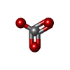
Mass: 18.015 Da / Num. of mol.: 8 / Source method: isolated from a natural source / Formula: H2O
-
Details
Has protein modification
Y
Sequence details
THE SEQUENCE OF THIS ISOFORM DIFFERS FROM THE CANONICAL SEQUENCE BY THE RESIDUES 994-1001 IN WHICH ...THE SEQUENCE OF THIS ISOFORM DIFFERS FROM THE CANONICAL SEQUENCE BY THE RESIDUES 994-1001 IN WHICH DPEDERRK IS REPLACED BY G
-
Experimental details
-
Experiment
Experiment
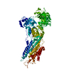
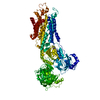
Method: X-RAY DIFFRACTION / Number of used crystals: 1
-
Sample preparation
Crystal
Density Matthews: 3.3 Å3/Da / Density % sol: 62.8 % / Description: NONE
Crystal grow
Temperature: 292 K / Method: vapor diffusion, hanging drop / pH: 6.8 Details: 19.5% (W/V) PEG2000-MME, 11% (V/V) GLYCEROL, 100 MM MGCL2, 3% (V/V) T-BUTANOL. THE CRYSTALLIZATION DROP WAS EQUILIBRATED AT 19 DEGREES C, AND THE CRYSTAL WAS FLASH FROZEN 13 DAYS AFTER SET-UP., pH 6.8
-
Data collection
Diffraction
Mean temperature: 100 K
Diffraction source
Source: SYNCHROTRON / Site: PETRA III, EMBL c/o DESY / Beamline: P14 (MX2) / Wavelength: 0.97626
In the structure databanks used in Yorodumi, some data are registered as the other names, "COVID-19 virus" and "2019-nCoV". Here are the details of the virus and the list of structure data.
Jan 31, 2019. EMDB accession codes are about to change! (news from PDBe EMDB page)
EMDB accession codes are about to change! (news from PDBe EMDB page)
The allocation of 4 digits for EMDB accession codes will soon come to an end. Whilst these codes will remain in use, new EMDB accession codes will include an additional digit and will expand incrementally as the available range of codes is exhausted. The current 4-digit format prefixed with “EMD-” (i.e. EMD-XXXX) will advance to a 5-digit format (i.e. EMD-XXXXX), and so on. It is currently estimated that the 4-digit codes will be depleted around Spring 2019, at which point the 5-digit format will come into force.
The EM Navigator/Yorodumi systems omit the EMD- prefix.
Related info.:Q: What is EMD? / ID/Accession-code notation in Yorodumi/EM Navigator
Yorodumi is a browser for structure data from EMDB, PDB, SASBDB, etc.
This page is also the successor to EM Navigator detail page, and also detail information page/front-end page for Omokage search.
The word "yorodu" (or yorozu) is an old Japanese word meaning "ten thousand". "mi" (miru) is to see.
Related info.:EMDB / PDB / SASBDB / Comparison of 3 databanks / Yorodumi Search / Aug 31, 2016. New EM Navigator & Yorodumi / Yorodumi Papers / Jmol/JSmol / Function and homology information / Changes in new EM Navigator and Yorodumi
 Movie
Movie Controller

 Yorodumi
 Open data
 Basic information
 Components
 Keywords
 Function and homology information
 X-RAY DIFFRACTION /
  Authors
 Citation
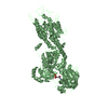
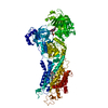
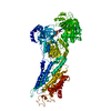
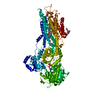
 Structure visualization
 Downloads & links
 Other downloads


 PDBj
PDBj

 Assembly
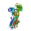
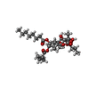


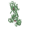
 Mass: 650.754 Da / Num. of mol.: 1 / Source method: obtained synthetically / Formula: C34H50O12
 Mass: 98.940 Da / Num. of mol.: 1 / Source method: obtained synthetically / Formula: VO3
 Mass: 715.289 Da / Num. of mol.: 1 / Source method: obtained synthetically / Formula: C17H18N8O18P3
 Mass: 24.305 Da / Num. of mol.: 2 / Source method: obtained synthetically / Formula: Mg
 Mass: 39.098 Da / Num. of mol.: 1 / Source method: obtained synthetically / Formula: K
 Mass: 35.453 Da / Num. of mol.: 1 / Source method: obtained synthetically / Formula: Cl
 Sample preparation
 / Beamline: P14 (MX2) / Wavelength: 0.97626
  Processing