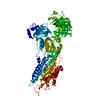
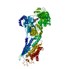
[English] 日本語
Yorodumi
- PDB-3n5k: Structure Of The (Sr)Ca2+-ATPase E2-AlF4- Form -

+
Open data


ID or keywords:

Loading...

-
Basic information

Entry
Database: PDB / ID: 3n5k
TitleStructure Of The (Sr)Ca2+-ATPase E2-AlF4- Form
ComponentsSarcoplasmic/endoplasmic reticulum calcium ATPase 1
KeywordsHYDROLASE / SERCA / Adenosine Triphosphate / Calcium-Transporting ATPases / Thapsigargin
Function / homology
Function and homology information


positive regulation of cardiac muscle cell contraction / positive regulation of calcium ion import into sarcoplasmic reticulum / H zone / positive regulation of fast-twitch skeletal muscle fiber contraction / calcium ion import into sarcoplasmic reticulum / regulation of striated muscle contraction / negative regulation of striated muscle contraction / positive regulation of ATPase-coupled calcium transmembrane transporter activity / P-type Ca2+ transporter / P-type calcium transporter activity ...positive regulation of cardiac muscle cell contraction / positive regulation of calcium ion import into sarcoplasmic reticulum / H zone / positive regulation of fast-twitch skeletal muscle fiber contraction / calcium ion import into sarcoplasmic reticulum / regulation of striated muscle contraction / negative regulation of striated muscle contraction / positive regulation of ATPase-coupled calcium transmembrane transporter activity / P-type Ca2+ transporter / P-type calcium transporter activity / I band / endoplasmic reticulum-Golgi intermediate compartment / sarcoplasmic reticulum membrane / sarcoplasmic reticulum / calcium ion transport / calcium ion binding / endoplasmic reticulum membrane / perinuclear region of cytoplasm / endoplasmic reticulum / ATP hydrolysis activity / ATP binding / membrane
Similarity search - Function
Calcium-transporting ATPase, transmembrane domain / Calcium-transporting ATPase, transmembrane domain / P-type ATPase, subfamily IIA, SERCA-type / Calcium-transporting ATPase, cytoplasmic transduction domain A / Calcium-transporting ATPase, cytoplasmic transduction domain A / Calcium-transporting ATPase, cytoplasmic domain N / Calcium-transporting ATPase, cytoplasmic domain N / haloacid dehalogenase-like hydrolase / Cation-transporting P-type ATPase, C-terminal / Cation transporting ATPase, C-terminus ...Calcium-transporting ATPase, transmembrane domain / Calcium-transporting ATPase, transmembrane domain / P-type ATPase, subfamily IIA, SERCA-type / Calcium-transporting ATPase, cytoplasmic transduction domain A / Calcium-transporting ATPase, cytoplasmic transduction domain A / Calcium-transporting ATPase, cytoplasmic domain N / Calcium-transporting ATPase, cytoplasmic domain N / haloacid dehalogenase-like hydrolase / Cation-transporting P-type ATPase, C-terminal / Cation transporting ATPase, C-terminus / Cation transporter/ATPase, N-terminus / Cation-transporting P-type ATPase, N-terminal / Cation transporter/ATPase, N-terminus / Cation transport ATPase (P-type) / E1-E2 ATPase / P-type ATPase, haloacid dehalogenase domain / P-type ATPase, phosphorylation site / P-type ATPase, cytoplasmic domain N / E1-E2 ATPases phosphorylation site. / P-type ATPase, A domain superfamily / P-type ATPase / P-type ATPase, transmembrane domain superfamily / haloacid dehalogenase-like hydrolase / HAD superfamily/HAD-like / HAD superfamily / HAD-like superfamily / Distorted Sandwich / Up-down Bundle / Rossmann fold / 3-Layer(aba) Sandwich / Mainly Beta / Mainly Alpha / Alpha Beta
Similarity search - Domain/homology
ACETATE ION / TETRAFLUOROALUMINATE ION / : / Chem-TG1 / Sarcoplasmic/endoplasmic reticulum calcium ATPase 1
Similarity search - Component
Biological speciesOryctolagus cuniculus (rabbit)
MethodX-RAY DIFFRACTION / SYNCHROTRON / MOLECULAR REPLACEMENT / Resolution: 2.2 Å
AuthorsBublitz, M. / Olesen, C. / Poulsen, H. / Morth, J.P. / Moller, J.V. / Nissen, P.
CitationJournal: J.Biol.Chem. / Year: 2013
Title: Ion pathways in the sarcoplasmic reticulum Ca2+-ATPase.
Authors: Bublitz, M. / Musgaard, M. / Poulsen, H. / Thogersen, L. / Olesen, C. / Schiott, B. / Morth, J.P. / Moller, J.V. / Nissen, P.
History
DepositionMay 25, 2010Deposition site: RCSB / Processing site: RCSB
Revision 1.0Jun 8, 2011Provider: repository / Type: Initial release
Revision 1.1Jul 13, 2011Group: Version format compliance
Revision 1.2Feb 27, 2013Group: Database references
Revision 1.3Mar 19, 2014Group: Refinement description
Revision 1.4Sep 6, 2023Group: Data collection / Database references ...Data collection / Database references / Derived calculations / Refinement description / Structure summary
Category: chem_comp / chem_comp_atom ...chem_comp / chem_comp_atom / chem_comp_bond / database_2 / entity / pdbx_entity_nonpoly / pdbx_initial_refinement_model / pdbx_struct_conn_angle / struct_conn / struct_site
Item: _chem_comp.name / _database_2.pdbx_DOI ..._chem_comp.name / _database_2.pdbx_DOI / _database_2.pdbx_database_accession / _entity.pdbx_description / _pdbx_entity_nonpoly.name / _pdbx_struct_conn_angle.ptnr1_auth_asym_id / _pdbx_struct_conn_angle.ptnr1_auth_comp_id / _pdbx_struct_conn_angle.ptnr1_auth_seq_id / _pdbx_struct_conn_angle.ptnr1_label_asym_id / _pdbx_struct_conn_angle.ptnr1_label_atom_id / _pdbx_struct_conn_angle.ptnr1_label_comp_id / _pdbx_struct_conn_angle.ptnr1_label_seq_id / _pdbx_struct_conn_angle.ptnr2_auth_asym_id / _pdbx_struct_conn_angle.ptnr2_auth_comp_id / _pdbx_struct_conn_angle.ptnr2_auth_seq_id / _pdbx_struct_conn_angle.ptnr2_label_asym_id / _pdbx_struct_conn_angle.ptnr2_label_atom_id / _pdbx_struct_conn_angle.ptnr2_label_comp_id / _pdbx_struct_conn_angle.ptnr3_auth_asym_id / _pdbx_struct_conn_angle.ptnr3_auth_comp_id / _pdbx_struct_conn_angle.ptnr3_auth_seq_id / _pdbx_struct_conn_angle.ptnr3_label_asym_id / _pdbx_struct_conn_angle.ptnr3_label_atom_id / _pdbx_struct_conn_angle.ptnr3_label_comp_id / _pdbx_struct_conn_angle.ptnr3_label_seq_id / _pdbx_struct_conn_angle.value / _struct_conn.pdbx_dist_value / _struct_conn.ptnr1_auth_asym_id / _struct_conn.ptnr1_auth_comp_id / _struct_conn.ptnr1_auth_seq_id / _struct_conn.ptnr1_label_asym_id / _struct_conn.ptnr1_label_atom_id / _struct_conn.ptnr1_label_comp_id / _struct_conn.ptnr1_label_seq_id / _struct_conn.ptnr2_auth_asym_id / _struct_conn.ptnr2_auth_comp_id / _struct_conn.ptnr2_auth_seq_id / _struct_conn.ptnr2_label_asym_id / _struct_conn.ptnr2_label_atom_id / _struct_conn.ptnr2_label_comp_id / _struct_site.pdbx_auth_asym_id / _struct_site.pdbx_auth_comp_id / _struct_site.pdbx_auth_seq_id

-
Structure visualization

Structure viewerMolecule:
MolmilJmol/JSmol

Downloads & links

-
Assembly

Deposited unit
A: Sarcoplasmic/endoplasmic reticulum calcium ATPase 1
B: Sarcoplasmic/endoplasmic reticulum calcium ATPase 1
hetero molecules


Theoretical massNumber of molelcules
Total (without water)221,07614
Polymers219,2052
Non-polymers1,87012
Water14,772820
1
A: Sarcoplasmic/endoplasmic reticulum calcium ATPase 1
hetero molecules


Theoretical massNumber of molelcules
Total (without water)110,5387
Polymers109,6031
Non-polymers9356
Water181
TypeNameSymmetry operationNumber
identity operation1_555x,y,z1
MethodPISA
2
B: Sarcoplasmic/endoplasmic reticulum calcium ATPase 1
hetero molecules


Theoretical massNumber of molelcules
Total (without water)110,5387
Polymers109,6031
Non-polymers9356
Water181
TypeNameSymmetry operationNumber
identity operation1_555x,y,z1
MethodPISA
Unit cell
Length a, b, c (Å)101.930, 109.420, 276.092
Angle α, β, γ (deg.)90.00, 90.00, 90.00
Int Tables number19
Space group name H-MP212121

-
Components

-
Protein , 1 types, 2 molecules AB

#1: Protein Sarcoplasmic/endoplasmic reticulum calcium ATPase 1 / SR Ca(2+)-ATPase 1 / SERCA1 / Calcium pump 1 / Calcium-transporting ATPase sarcoplasmic reticulum ...SR Ca(2+)-ATPase 1 / SERCA1 / Calcium pump 1 / Calcium-transporting ATPase sarcoplasmic reticulum type / fast twitch skeletal muscle isoform / Endoplasmic reticulum class 1/2 Ca(2+) ATPase


Mass: 109602.578 Da / Num. of mol.: 2 / Source method: isolated from a natural source / Source: (natural) Oryctolagus cuniculus (rabbit) / References: UniProt: P04191, EC: 3.6.3.8

-
Non-polymers , 6 types, 832 molecules

#2: Chemical ChemComp-TG1 / OCTANOIC ACID [3S-[3ALPHA, 3ABETA, 4ALPHA, 6BETA, 6ABETA, 7BETA, 8ALPHA(Z), 9BALPHA]]-6-(ACETYLOXY)-2,3,-3A,4,5,6,6A,7,8,9B-DECAHYDRO-3,3A-DIHYDROXY-3,6,9-TRIMETHYL-8-[(2-METHYL-1-OXO-2-BUTENYL)OX Y]-2-OXO-4-(1-OXOBUTOXY)-AZULENO[4,5-B]FURAN-7-YL ESTER / THAPSIGARGIN / Thapsigargin


Mass: 650.754 Da / Num. of mol.: 2 / Source method: obtained synthetically / Formula: C34H50O12 / Comment: inhibitor*YM
#3: Chemical ChemComp-MG / MAGNESIUM ION


Mass: 24.305 Da / Num. of mol.: 2 / Source method: obtained synthetically / Formula: Mg
#4: Chemical ChemComp-ALF / TETRAFLUOROALUMINATE ION


Mass: 102.975 Da / Num. of mol.: 2 / Source method: obtained synthetically / Formula: AlF4
#5: Chemical ChemComp-K / POTASSIUM ION


Mass: 39.098 Da / Num. of mol.: 2 / Source method: obtained synthetically / Formula: K
#6: Chemical
ChemComp-ACT / ACETATE ION / Acetate


Mass: 59.044 Da / Num. of mol.: 4 / Source method: obtained synthetically / Formula: C2H3O2
#7: Water ChemComp-HOH / water / Water


Mass: 18.015 Da / Num. of mol.: 820 / Source method: isolated from a natural source / Formula: H2O

-
Details

Sequence detailsRESIDUES DPEDERRK (994-1001) HAVE BEEN REPLACED BY G IN ISOFORM P04191-2

-
Experimental details

-
Experiment

ExperimentMethod: X-RAY DIFFRACTION / Number of used crystals: 1

-
Sample preparation

CrystalDensity Matthews: 3.48 Å3/Da / Density % sol: 64.67 %
Crystal growTemperature: 290 K / Method: vapor diffusion, hanging drop / pH: 6.6
Details: 13% PEG 6000, 6% 2-methyl-2,4-pentane diol, 70mM sodium acetate, pH 6.6, VAPOR DIFFUSION, HANGING DROP, temperature 290K

-
Data collection

DiffractionMean temperature: 100 K
Diffraction sourceSource: SYNCHROTRON / Site: SLS / Beamline: X06SA / Wavelength: 1.0007 Å
DetectorType: PSI PILATUS 6M / Detector: PIXEL / Date: Oct 11, 2009
RadiationMonochromator: LN2 cooled fixed-exit Si(111) monochromator / Protocol: SINGLE WAVELENGTH / Monochromatic (M) / Laue (L): M / Scattering type: x-ray
Radiation wavelengthWavelength: 1.0007 Å / Relative weight: 1
ReflectionResolution: 2.2→82 Å / Num. obs: 156560 / % possible obs: 99.8 % / Observed criterion σ(F): 0 / Observed criterion σ(I): 0 / Redundancy: 10.1 % / Biso Wilson estimate: 38.7 Å2 / Rmerge(I) obs: 0.117 / Net I/σ(I): 14.2
Reflection shellResolution: 2.2→2.32 Å / Redundancy: 9.5 % / Mean I/σ(I) obs: 2.5 / % possible all: 99.6

-
Processing

Software
NameVersionClassification
remdaq.pilatusdata collection
PHASERphasing
PHENIX(phenix.refine: 1.6.1_357)refinement
MOSFLMdata reduction
SCALAdata scaling
RefinementMethod to determine structure: MOLECULAR REPLACEMENT
Starting model: PDB entry 3FGO
Resolution: 2.2→72 Å / SU ML: 0.3 / Isotropic thermal model: anisotropic / σ(F): 1.34 / Stereochemistry target values: ML
RfactorNum. reflection% reflection
Rfree0.216 7823 5 %
Rwork0.184 --
obs0.186 156449 99.7 %
Solvent computationShrinkage radii: 0.9 Å / VDW probe radii: 1.11 Å / Solvent model: FLAT BULK SOLVENT MODEL / Bsol: 42.33 Å2 / ksol: 0.3 e/Å3
Displacement parametersBiso mean: 37.54 Å2
Baniso -1Baniso -2Baniso -3
1--3.6891 Å20 Å2-0 Å2
2--8.5793 Å2-0 Å2
3----4.8901 Å2
Refinement stepCycle: LAST / Resolution: 2.2→72 Å
ProteinNucleic acidLigandSolventTotal
Num. atoms15342 0 122 820 16284
Refine LS restraints
Refine-IDTypeDev idealNumber
X-RAY DIFFRACTIONf_bond_d0.0115742
X-RAY DIFFRACTIONf_angle_d1.17921356
X-RAY DIFFRACTIONf_dihedral_angle_d13.4555998
X-RAY DIFFRACTIONf_chiral_restr0.0792474
X-RAY DIFFRACTIONf_plane_restr0.0052720
LS refinement shell
Resolution (Å)Rfactor RfreeNum. reflection RfreeRfactor RworkNum. reflection RworkRefine-ID% reflection obs (%)
2.2-2.2250.26962540.23794889X-RAY DIFFRACTION100
2.225-2.25120.31222650.27324851X-RAY DIFFRACTION99
2.2512-2.27860.23712530.2344903X-RAY DIFFRACTION99
2.2786-2.30750.24282600.21484892X-RAY DIFFRACTION99
2.3075-2.33790.25072600.2144944X-RAY DIFFRACTION100
2.3379-2.36990.24692560.21044912X-RAY DIFFRACTION100
2.3699-2.40370.27262510.20454808X-RAY DIFFRACTION99
2.4037-2.43960.21042590.19814940X-RAY DIFFRACTION100
2.4396-2.47770.23562620.19314912X-RAY DIFFRACTION100
2.4777-2.51840.25992540.19274905X-RAY DIFFRACTION100
2.5184-2.56180.23672580.1824931X-RAY DIFFRACTION100
2.5618-2.60840.22962630.17764939X-RAY DIFFRACTION100
2.6084-2.65860.23792600.17354910X-RAY DIFFRACTION100
2.6586-2.71280.20432620.1794927X-RAY DIFFRACTION100
2.7128-2.77180.19712590.16834951X-RAY DIFFRACTION100
2.7718-2.83630.20382540.16954922X-RAY DIFFRACTION100
2.8363-2.90720.21682640.17844937X-RAY DIFFRACTION100
2.9072-2.98580.21462580.17594925X-RAY DIFFRACTION100
2.9858-3.07370.22872610.17814925X-RAY DIFFRACTION100
3.0737-3.17290.2032600.17835001X-RAY DIFFRACTION100
3.1729-3.28630.22542630.17924948X-RAY DIFFRACTION100
3.2863-3.41790.20692630.18464997X-RAY DIFFRACTION100
3.4179-3.57340.20782590.17744961X-RAY DIFFRACTION100
3.5734-3.76180.20582670.17294996X-RAY DIFFRACTION100
3.7618-3.99750.17842610.16414954X-RAY DIFFRACTION100
3.9975-4.30610.18532630.15665020X-RAY DIFFRACTION100
4.3061-4.73940.20872660.1545036X-RAY DIFFRACTION100
4.7394-5.4250.18972650.16545054X-RAY DIFFRACTION100
5.425-6.8340.20562680.18835103X-RAY DIFFRACTION99
6.834-72.04050.19282750.1795233X-RAY DIFFRACTION98
Refinement TLS params.

Method: refined / Refine-ID: X-RAY DIFFRACTION

IDL112)L122)L132)L222)L232)L332)S11 (Å °)S12 (Å °)S13 (Å °)S21 (Å °)S22 (Å °)S23 (Å °)S31 (Å °)S32 (Å °)S33 (Å °)T112)T122)T132)T222)T232)T332)Origin x (Å)Origin y (Å)Origin z (Å)
10.3817-0.4941-0.14420.51120.17940.09520.24290.4516-0.0452-0.6666-0.23070.16480.1973-0.3546-0.00530.89860.0427-0.15020.80.07060.256536.189156.97399.1808
21.33610.0684-0.03061.15550.14390.04240.04970.78890.0996-0.87430.0493-0.0359-0.5544-0.1524-0.0891.10150.11010.03490.72710.2380.180946.599374.54261.1783
30.83940.04080.18670.37250.2190.4045-0.0131-0.007-0.00540.00780.01260.00830.0199-0.01850.00440.2002-0.01250.02190.08140.01640.211154.638349.099260.6381
40.0304-0.0361-0.11410.04560.1880.58880.01680.31810.0118-0.2915-0.04140.0427-0.326-0.29980.04190.74640.03570.05010.64480.00690.242210.889217.4664.7501
50.5602-0.1003-0.27930.67210.25540.2046-0.02540.04580.0125-0.3333-0.0364-0.1280.09830.70290.06230.6067-0.00490.14380.76380.01950.22529.72636.91951.5198
60.983-0.0517-0.33490.6361-0.30960.6773-0.0291-0.0303-0.0749-0.0395-0.02150.08360.00730.05670.04640.12140.0164-0.02020.1072-0.02270.1637-5.43317.123456.2834
Refinement TLS group
IDRefine-IDRefine TLS-IDSelection detailsAuth asym-IDAuth seq-ID
1X-RAY DIFFRACTION1( CHAIN A AND ( RESID 45:122 OR RESID 238:329 ) )A45 - 122
2X-RAY DIFFRACTION1( CHAIN A AND ( RESID 45:122 OR RESID 238:329 ) )A238 - 329
3X-RAY DIFFRACTION2( CHAIN A AND RESID 742:994 )A742 - 994
4X-RAY DIFFRACTION3( CHAIN A AND ( RESID 1:44 OR RESID 123:237 ) )A1 - 44
5X-RAY DIFFRACTION3( CHAIN A AND ( RESID 1:44 OR RESID 123:237 ) )A123 - 237
6X-RAY DIFFRACTION4( CHAIN B AND ( RESID 45:122 OR RESID 238:329 ) )B45 - 122
7X-RAY DIFFRACTION4( CHAIN B AND ( RESID 45:122 OR RESID 238:329 ) )B238 - 329
8X-RAY DIFFRACTION5( CHAIN B AND RESID 742:994 )B742 - 994
9X-RAY DIFFRACTION6( CHAIN B AND ( RESID 1:44 OR RESID 123:237 ) )B1 - 44
10X-RAY DIFFRACTION6( CHAIN B AND ( RESID 1:44 OR RESID 123:237 ) )B123 - 237

+
About Yorodumi

-
News

-
Feb 9, 2022. New format data for meta-information of EMDB entries

New format data for meta-information of EMDB entries

  • Version 3 of the EMDB header file is now the official format.
  • The previous official version 1.9 will be removed from the archive.

Related info.:EMDB header

External links:wwPDB to switch to version 3 of the EMDB data model

-
Aug 12, 2020. Covid-19 info

Covid-19 info

URL: https://pdbj.org/emnavi/covid19.php

New page: Covid-19 featured information page in EM Navigator.

Related info.:Covid-19 info / Mar 5, 2020. Novel coronavirus structure data

+
Mar 5, 2020. Novel coronavirus structure data

Novel coronavirus structure data

Related info.:Yorodumi Speices / Aug 12, 2020. Covid-19 info

External links:COVID-19 featured content - PDBj / Molecule of the Month (242):Coronavirus Proteases

+
Jan 31, 2019. EMDB accession codes are about to change! (news from PDBe EMDB page)

EMDB accession codes are about to change! (news from PDBe EMDB page)

  • The allocation of 4 digits for EMDB accession codes will soon come to an end. Whilst these codes will remain in use, new EMDB accession codes will include an additional digit and will expand incrementally as the available range of codes is exhausted. The current 4-digit format prefixed with “EMD-” (i.e. EMD-XXXX) will advance to a 5-digit format (i.e. EMD-XXXXX), and so on. It is currently estimated that the 4-digit codes will be depleted around Spring 2019, at which point the 5-digit format will come into force.
  • The EM Navigator/Yorodumi systems omit the EMD- prefix.

Related info.:Q: What is EMD? / ID/Accession-code notation in Yorodumi/EM Navigator

External links:EMDB Accession Codes are Changing Soon! / Contact to PDBj

+
Jul 12, 2017. Major update of PDB

Major update of PDB

  • wwPDB released updated PDB data conforming to the new PDBx/mmCIF dictionary.
  • This is a major update changing the version number from 4 to 5, and with Remediation, in which all the entries are updated.
  • In this update, many items about electron microscopy experimental information are reorganized (e.g. em_software).
  • Now, EM Navigator and Yorodumi are based on the updated data.

External links:wwPDB Remediation / Enriched Model Files Conforming to OneDep Data Standards Now Available in the PDB FTP Archive

-
Yorodumi

Thousand views of thousand structures

  • Yorodumi is a browser for structure data from EMDB, PDB, SASBDB, etc.
  • This page is also the successor to EM Navigator detail page, and also detail information page/front-end page for Omokage search.
  • The word "yorodu" (or yorozu) is an old Japanese word meaning "ten thousand". "mi" (miru) is to see.

Related info.:EMDB / PDB / SASBDB / Comparison of 3 databanks / Yorodumi Search / Aug 31, 2016. New EM Navigator & Yorodumi / Yorodumi Papers / Jmol/JSmol / Function and homology information / Changes in new EM Navigator and Yorodumi

Read more