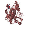
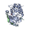
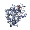
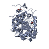
Entry Database : PDB / ID : 4zv7Title Crystal structure of hexagonal form of lipase B from Candida antarctica Lipase B Keywords / / Function / homology Function Domain/homology Component
Biological species Candida antarctica (fungus)Method / / Resolution : 2 Å Authors Strzelczyk, P. / Blaszczyk, J. / Bujacz, G. Journal : Acta Biochim.Pol. / Year : 2016Title : Crystal and molecular structure of hexagonal form of lipase B from Candida antarctica.Authors : Strzelczyk, P. / Bujacz, G.D. / Kiebasinski, P. / Baszczyk, J. History Deposition May 18, 2015 Deposition site / Processing site Revision 1.0 Jan 13, 2016 Provider / Type Revision 1.1 Mar 30, 2016 Group Revision 1.2 Sep 13, 2017 Group / Category / Item Revision 2.0 Jul 29, 2020 Group Advisory / Atomic model ... Advisory / Atomic model / Data collection / Derived calculations / Structure summary Category atom_site / chem_comp ... atom_site / chem_comp / entity / pdbx_branch_scheme / pdbx_chem_comp_identifier / pdbx_entity_branch / pdbx_entity_branch_descriptor / pdbx_entity_branch_link / pdbx_entity_branch_list / pdbx_entity_nonpoly / pdbx_nonpoly_scheme / pdbx_struct_assembly_gen / pdbx_validate_close_contact / struct_asym / struct_conn / struct_site / struct_site_gen Item _atom_site.auth_asym_id / _atom_site.auth_seq_id ... _atom_site.auth_asym_id / _atom_site.auth_seq_id / _atom_site.label_asym_id / _chem_comp.name / _chem_comp.type / _entity.formula_weight / _entity.pdbx_description / _entity.pdbx_number_of_molecules / _entity.type / _pdbx_struct_assembly_gen.asym_id_list / _pdbx_validate_close_contact.auth_asym_id_2 / _pdbx_validate_close_contact.auth_seq_id_2 / _struct_conn.pdbx_role / _struct_conn.ptnr1_auth_asym_id / _struct_conn.ptnr1_auth_seq_id / _struct_conn.ptnr2_auth_asym_id / _struct_conn.ptnr2_auth_seq_id / _struct_conn.ptnr2_label_asym_id Description / Provider / Type Revision 2.1 Jan 10, 2024 Group Data collection / Database references ... Data collection / Database references / Refinement description / Structure summary Category chem_comp / chem_comp_atom ... chem_comp / chem_comp_atom / chem_comp_bond / database_2 / pdbx_initial_refinement_model Item / _database_2.pdbx_DOI / _database_2.pdbx_database_accessionRevision 2.2 Nov 13, 2024 Group / Category / pdbx_modification_feature
Show all Show less
 Movie
Movie Controller

 Yorodumi
 Open data
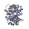
 Basic information
 Components
 Keywords
 Function and homology information
 Candida antarctica (fungus)
 X-RAY DIFFRACTION /
  Authors
 Citation
 Structure visualization
 Downloads & links
 Other downloads
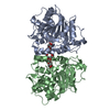

 PDBj
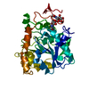
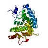
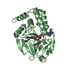
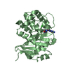
PDBj Assembly
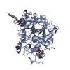
 Mass: 18.015 Da / Num. of mol.: 332 / Source method: isolated from a natural source / Formula: H2O
 Sample preparation
 Processing