 Movie
Movie Controller

[English] 日本語
 Yorodumi
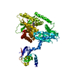
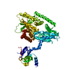
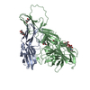
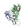
- PDB-4z94: Actin Complex With a Chimera of Tropomodulin-1 and Leiomodin-1 Ac... -
+ Open data

- Basic information
| Entry | Database: PDB / ID: 4z94 | ||||||
|---|---|---|---|---|---|---|---|
| Title | Actin Complex With a Chimera of Tropomodulin-1 and Leiomodin-1 Actin-Binding Site 2 | ||||||
 Components Components |
| ||||||
 Keywords Keywords | PROTEIN BINDING/STRUCTURAL PROTEIN / Actin Nucleator / ATP-actin / PROTEIN BINDING-STRUCTURAL PROTEIN complex | ||||||
| Function / homology |  Function and homology information Function and homology informationpointed-end actin filament capping / lens fiber cell development / actin nucleation / striated muscle atrophy / regulation of establishment of T cell polarity / regulation of plasma membrane raft polarization / regulation of receptor clustering / positive regulation of keratinocyte apoptotic process / renal protein absorption / positive regulation of protein processing in phagocytic vesicle ...pointed-end actin filament capping / lens fiber cell development / actin nucleation / striated muscle atrophy / regulation of establishment of T cell polarity / regulation of plasma membrane raft polarization / regulation of receptor clustering / positive regulation of keratinocyte apoptotic process / renal protein absorption / positive regulation of protein processing in phagocytic vesicle / phosphatidylinositol 3-kinase catalytic subunit binding / positive regulation of actin nucleation / myofibril assembly / actin cap / myosin II binding / host-mediated suppression of symbiont invasion / actin filament severing / barbed-end actin filament capping / cell projection assembly / actin polymerization or depolymerization / actin filament depolymerization / Striated Muscle Contraction / actin filament capping / relaxation of cardiac muscle / Sensory processing of sound by outer hair cells of the cochlea / phagocytosis, engulfment / cardiac muscle cell contraction / cytoskeletal motor activator activity / myosin heavy chain binding / hepatocyte apoptotic process / tropomyosin binding / actin filament bundle / troponin I binding / filamentous actin / mesenchyme migration / positive regulation of actin filament polymerization / cortical cytoskeleton / myofibril / skeletal muscle myofibril / sarcoplasm / actin filament bundle assembly / striated muscle thin filament / skeletal muscle thin filament assembly / cilium assembly / actin monomer binding / Caspase-mediated cleavage of cytoskeletal proteins / Smooth Muscle Contraction / skeletal muscle fiber development / phagocytic vesicle / response to muscle stretch / stress fiber / titin binding / actin filament polymerization / phosphatidylinositol-4,5-bisphosphate binding / muscle contraction / actin filament organization / central nervous system development / sarcomere / filopodium / actin filament / adult locomotory behavior / protein destabilization / cellular response to type II interferon / Hydrolases; Acting on acid anhydrides; Acting on acid anhydrides to facilitate cellular and subcellular movement / calcium-dependent protein binding / actin filament binding / lamellipodium / actin cytoskeleton / actin binding / cell body / secretory granule lumen / blood microparticle / amyloid fibril formation / ficolin-1-rich granule lumen / cytoskeleton / Amyloid fiber formation / protein domain specific binding / focal adhesion / hydrolase activity / calcium ion binding / Neutrophil degranulation / positive regulation of gene expression / magnesium ion binding / : / extracellular exosome / extracellular region / ATP binding / membrane / identical protein binding / plasma membrane / cytoplasm / cytosol Similarity search - Function | ||||||
| Biological species |  Homo sapiens (human) Homo sapiens (human) | ||||||
| Method |  X-RAY DIFFRACTION / MOLECULAR REPLACEMENT / Resolution: 2.4 Å X-RAY DIFFRACTION / MOLECULAR REPLACEMENT / Resolution: 2.4 Å | ||||||
 Authors Authors | Rebowski, G. / Boczkowska, M. / Dominguez, R. | ||||||
| Funding support |  United States, 1items United States, 1items
| ||||||
 Citation Citation | Journal: Nat Commun / Year: 2015 Title: How Leiomodin and Tropomodulin use a common fold for different actin assembly functions. Authors: Boczkowska, M. / Rebowski, G. / Kremneva, E. / Lappalainen, P. / Dominguez, R. | ||||||
| History |
|
- Structure visualization
| Structure viewer | Molecule: MolmilJmol/JSmol |
|---|
- Downloads & links
-Download
| PDBx/mmCIF format | 4z94.cif.gz | 399.9 KB | Display | PDBx/mmCIF format |
|---|---|---|---|---|
| PDB format | pdb4z94.ent.gz | 326 KB | Display | PDB format |
| PDBx/mmJSON format | 4z94.json.gz | Tree view | PDBx/mmJSON format | |
| Others |  Other downloads Other downloads |
-Validation report
| Arichive directory | https://data.pdbj.org/pub/pdb/validation_reports/z9/4z94ftp://data.pdbj.org/pub/pdb/validation_reports/z9/4z94 | HTTPS FTP |
|---|
-Related structure data
| Related structure data | 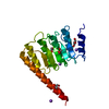 4z79C  4z8gC  4pkiS C: citing same article ( S: Starting model for refinement |
|---|---|
| Similar structure data |


-Links
 PDBj
PDBj


- Assembly
| Deposited unit | 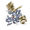
| ||||||||
|---|---|---|---|---|---|---|---|---|---|
| 1 |
| ||||||||
| Unit cell |
|
-Components
| #1: Protein | Mass: 42109.973 Da / Num. of mol.: 1 / Source method: isolated from a natural source / Source: (natural) | ||
|---|---|---|---|
| #2: Protein | Mass: 36533.457 Da / Num. of mol.: 1 Fragment: Gelsolin residues 12-136 (UNP), linker,Tropomodulin-1 residues 160-228 (UNP), Leiomodin-1 Actin-Binding Site 2 (UNP residues 364-486) Source method: isolated from a genetically manipulated source Source: (gene. exp.) Homo sapiens (human) / Gene: GSN, TMOD1, D9S57E, TMOD, LMOD1 / Production host:  References: UniProt: P06396, UniProt: P28289, UniProt: P29536 | ||
| #3: Chemical | ChemComp-ATP /   Mass: 507.181 Da / Num. of mol.: 1 / Source method: isolated from a natural source / Formula: C10H16N5O13P3 / Comment: ATP, energy-carrying molecule*YM Mass: 507.181 Da / Num. of mol.: 1 / Source method: isolated from a natural source / Formula: C10H16N5O13P3 / Comment: ATP, energy-carrying molecule*YM | ||
| #4: Chemical |   Mass: 40.078 Da / Num. of mol.: 3 / Source method: obtained synthetically / Formula: Ca Mass: 40.078 Da / Num. of mol.: 3 / Source method: obtained synthetically / Formula: Ca#5: Water | ChemComp-HOH / |  Mass: 18.015 Da / Num. of mol.: 182 / Source method: isolated from a natural source / Formula: H2O Mass: 18.015 Da / Num. of mol.: 182 / Source method: isolated from a natural source / Formula: H2O |
-Experimental details
-Experiment
| Experiment | Method: X-RAY DIFFRACTION |
|---|
- Sample preparation
| Crystal | Density Matthews: 2.62 Å3/Da / Density % sol: 53.08 % |
|---|---|
| Crystal grow | Temperature: 291.5 K / Method: vapor diffusion, hanging drop / pH: 8.8 Details: 20% PEG3350, 200 mM lithium sulfate, 100 mM Tris, pH 8.8, 15% glycerol |
-Data collection
| Diffraction | Mean temperature: 100 K |
|---|---|
| Diffraction source | Source: ROTATING ANODE / Type: BRUKER AXS MICROSTAR / Wavelength: 1.5418 Å |
| Detector | Type: APEX II CCD / Detector: CCD / Date: Feb 2, 2015 |
| Radiation | Protocol: SINGLE WAVELENGTH / Monochromatic (M) / Laue (L): M / Scattering type: x-ray |
| Radiation wavelength | Wavelength: 1.5418 Å / Relative weight: 1 |
| Reflection | Resolution: 2.4→39.9 Å / Num. all: 27926 / Num. obs: 27914 / % possible obs: 89.2 % / Redundancy: 4.8 % / Rmerge(I) obs: 0.127 / Net I/σ(I): 9 |
| Reflection shell | Resolution: 2.4→2.49 Å / Redundancy: 1.8 % / Rmerge(I) obs: 0.49 / Mean I/σ(I) obs: 1.92 / % possible all: 50.1 |
- Processing
| Software |
| ||||||||||||||||||||||||||||||||||||||||||||||||||||||||||||||||||||||||||||||||||||||||||||||||||||||||||||||||||||||||||||||||||||||||||||||||||||||
|---|---|---|---|---|---|---|---|---|---|---|---|---|---|---|---|---|---|---|---|---|---|---|---|---|---|---|---|---|---|---|---|---|---|---|---|---|---|---|---|---|---|---|---|---|---|---|---|---|---|---|---|---|---|---|---|---|---|---|---|---|---|---|---|---|---|---|---|---|---|---|---|---|---|---|---|---|---|---|---|---|---|---|---|---|---|---|---|---|---|---|---|---|---|---|---|---|---|---|---|---|---|---|---|---|---|---|---|---|---|---|---|---|---|---|---|---|---|---|---|---|---|---|---|---|---|---|---|---|---|---|---|---|---|---|---|---|---|---|---|---|---|---|---|---|---|---|---|---|---|---|---|
| Refinement | Method to determine structure: MOLECULAR REPLACEMENT Starting model: PDB entry 4PKI Resolution: 2.4→39.812 Å / SU ML: 0.3 / Cross valid method: FREE R-VALUE / σ(F): 1.33 / Phase error: 27.24 / Stereochemistry target values: ML
| ||||||||||||||||||||||||||||||||||||||||||||||||||||||||||||||||||||||||||||||||||||||||||||||||||||||||||||||||||||||||||||||||||||||||||||||||||||||
| Solvent computation | Shrinkage radii: 0.9 Å / VDW probe radii: 1.11 Å / Solvent model: FLAT BULK SOLVENT MODEL | ||||||||||||||||||||||||||||||||||||||||||||||||||||||||||||||||||||||||||||||||||||||||||||||||||||||||||||||||||||||||||||||||||||||||||||||||||||||
| Refinement step | Cycle: LAST / Resolution: 2.4→39.812 Å
| ||||||||||||||||||||||||||||||||||||||||||||||||||||||||||||||||||||||||||||||||||||||||||||||||||||||||||||||||||||||||||||||||||||||||||||||||||||||
| Refine LS restraints |
| ||||||||||||||||||||||||||||||||||||||||||||||||||||||||||||||||||||||||||||||||||||||||||||||||||||||||||||||||||||||||||||||||||||||||||||||||||||||
| LS refinement shell |
| ||||||||||||||||||||||||||||||||||||||||||||||||||||||||||||||||||||||||||||||||||||||||||||||||||||||||||||||||||||||||||||||||||||||||||||||||||||||
| Refinement TLS params. | Method: refined / Refine-ID: X-RAY DIFFRACTION
| ||||||||||||||||||||||||||||||||||||||||||||||||||||||||||||||||||||||||||||||||||||||||||||||||||||||||||||||||||||||||||||||||||||||||||||||||||||||
| Refinement TLS group |
|
