[English] 日本語
Yorodumi
- PDB-4pki: Complex of ATP-actin With the C-terminal Actin-Binding Domain of ... -

+
Open data


ID or keywords:

Loading...

-
Basic information

Entry
Database: PDB / ID: 4pki
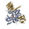
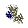
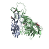
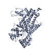
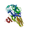
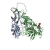
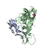
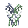
TitleComplex of ATP-actin With the C-terminal Actin-Binding Domain of Tropomodulin
Components
  • Actin, alpha skeletal muscle
  • Gelsolin,Tropomodulin-1 chimera
KeywordsContractile Protein/Actin-Binding Protein / Tmod / Actin Filament / Pointed-End Capping Protein / Tropomyosin / Contractile Protein / Actin-binding Protein / Contractile Protein-Actin-Binding Protein complex
Function / homology
Function and homology information


pointed-end actin filament capping / lens fiber cell development / myofibril assembly / striated muscle atrophy / regulation of establishment of T cell polarity / regulation of plasma membrane raft polarization / regulation of receptor clustering / renal protein absorption / positive regulation of keratinocyte apoptotic process / positive regulation of protein processing in phagocytic vesicle ...pointed-end actin filament capping / lens fiber cell development / myofibril assembly / striated muscle atrophy / regulation of establishment of T cell polarity / regulation of plasma membrane raft polarization / regulation of receptor clustering / renal protein absorption / positive regulation of keratinocyte apoptotic process / positive regulation of protein processing in phagocytic vesicle / positive regulation of actin nucleation / phosphatidylinositol 3-kinase catalytic subunit binding / positive regulation of cysteine-type endopeptidase activity involved in apoptotic signaling pathway / actin cap / sequestering of actin monomers / regulation of podosome assembly / myosin II binding / negative regulation of viral entry into host cell / actin filament severing / actin filament capping / barbed-end actin filament capping / actin filament depolymerization / actin polymerization or depolymerization / cell projection assembly / cardiac muscle cell contraction / Striated Muscle Contraction / podosome / sarcoplasm / Sensory processing of sound by outer hair cells of the cochlea / relaxation of cardiac muscle / cytoskeletal motor activator activity / phagocytosis, engulfment / cortical actin cytoskeleton / tropomyosin binding / cortical cytoskeleton / myofibril / myosin heavy chain binding / mesenchyme migration / troponin I binding / hepatocyte apoptotic process / actin filament bundle / filamentous actin / actin filament bundle assembly / skeletal muscle thin filament assembly / striated muscle thin filament / skeletal muscle myofibril / cilium assembly / actin monomer binding / Caspase-mediated cleavage of cytoskeletal proteins / skeletal muscle fiber development / phagocytic vesicle / stress fiber / titin binding / phosphatidylinositol-4,5-bisphosphate binding / response to muscle stretch / actin filament polymerization / sarcomere / adult locomotory behavior / filopodium / muscle contraction / central nervous system development / actin filament organization / actin filament / Hydrolases; Acting on acid anhydrides; Acting on acid anhydrides to facilitate cellular and subcellular movement / protein destabilization / cellular response to type II interferon / calcium-dependent protein binding / actin filament binding / actin cytoskeleton / lamellipodium / cell body / actin binding / blood microparticle / secretory granule lumen / ficolin-1-rich granule lumen / amyloid fibril formation / cytoskeleton / hydrolase activity / Amyloid fiber formation / protein domain specific binding / focal adhesion / calcium ion binding / Neutrophil degranulation / positive regulation of gene expression / magnesium ion binding / extracellular space / extracellular exosome / extracellular region / ATP binding / membrane / identical protein binding / plasma membrane / cytosol / cytoplasm
Similarity search - Function
Tropomodulin / Tropomodulin / Actin; Chain A, domain 2 / Actin; Chain A, domain 2 / Villin/Gelsolin / Gelsolin homology domain / Severin / Severin / Gelsolin-like domain / Gelsolin repeat ...Tropomodulin / Tropomodulin / Actin; Chain A, domain 2 / Actin; Chain A, domain 2 / Villin/Gelsolin / Gelsolin homology domain / Severin / Severin / Gelsolin-like domain / Gelsolin repeat / ADF-H/Gelsolin-like domain superfamily / Leucine-rich repeat, LRR (right-handed beta-alpha superhelix) / Ribonuclease Inhibitor / ATPase, substrate binding domain, subdomain 4 / Actin; Chain A, domain 4 / Alpha-Beta Horseshoe / ATPase, nucleotide binding domain / Actins signature 1. / Actin, conserved site / Actins signature 2. / Actin/actin-like conserved site / Actins and actin-related proteins signature. / Actin / Actin family / Actin / Leucine-rich repeat domain superfamily / ATPase, nucleotide binding domain / Nucleotidyltransferase; domain 5 / Roll / Alpha-Beta Complex / 2-Layer Sandwich / 3-Layer(aba) Sandwich / Mainly Beta / Alpha Beta
Similarity search - Domain/homology
ADENOSINE-5'-TRIPHOSPHATE / Gelsolin / Tropomodulin-1 / Actin, alpha skeletal muscle
Similarity search - Component
Biological speciesHomo sapiens (human)
Oryctolagus cuniculus (rabbit)
MethodX-RAY DIFFRACTION / SYNCHROTRON / MOLECULAR REPLACEMENT / Resolution: 2.3 Å
AuthorsRao, J.N. / Dominguez, R.
Funding support United States, 1items
OrganizationGrant numberCountry
National Institutes of Health/National Institute of General Medical Sciences (NIH/NIGMS)R01 GM073791 United States
CitationJournal: Science / Year: 2014
Title: Actin cytoskeleton. Mechanism of actin filament pointed-end capping by tropomodulin.
Authors: Rao, J.N. / Madasu, Y. / Dominguez, R.
History
DepositionMay 14, 2014Deposition site: RCSB / Processing site: RCSB
Revision 1.0Jul 30, 2014Provider: repository / Type: Initial release
Revision 1.1Aug 6, 2014Group: Database references
Revision 1.2Sep 6, 2017Group: Advisory / Author supporting evidence ...Advisory / Author supporting evidence / Database references / Derived calculations / Other / Source and taxonomy
Category: citation / entity_src_gen ...citation / entity_src_gen / entity_src_nat / pdbx_audit_support / pdbx_database_status / pdbx_struct_oper_list / pdbx_validate_close_contact
Item: _citation.journal_id_CSD / _entity_src_gen.pdbx_alt_source_flag ..._citation.journal_id_CSD / _entity_src_gen.pdbx_alt_source_flag / _entity_src_nat.pdbx_alt_source_flag / _pdbx_audit_support.funding_organization / _pdbx_database_status.pdb_format_compatible / _pdbx_struct_oper_list.symmetry_operation
Revision 1.3Dec 25, 2019Group: Author supporting evidence / Category: pdbx_audit_support / Item: _pdbx_audit_support.funding_organization
Revision 1.4Sep 27, 2023Group: Data collection / Database references ...Data collection / Database references / Derived calculations / Refinement description
Category: chem_comp_atom / chem_comp_bond ...chem_comp_atom / chem_comp_bond / database_2 / pdbx_initial_refinement_model / pdbx_struct_conn_angle / refine_hist / struct_conn
Item: _database_2.pdbx_DOI / _database_2.pdbx_database_accession ..._database_2.pdbx_DOI / _database_2.pdbx_database_accession / _pdbx_struct_conn_angle.ptnr1_auth_asym_id / _pdbx_struct_conn_angle.ptnr1_auth_comp_id / _pdbx_struct_conn_angle.ptnr1_auth_seq_id / _pdbx_struct_conn_angle.ptnr1_label_asym_id / _pdbx_struct_conn_angle.ptnr1_label_atom_id / _pdbx_struct_conn_angle.ptnr1_label_comp_id / _pdbx_struct_conn_angle.ptnr1_label_seq_id / _pdbx_struct_conn_angle.ptnr2_auth_asym_id / _pdbx_struct_conn_angle.ptnr2_auth_seq_id / _pdbx_struct_conn_angle.ptnr2_label_asym_id / _pdbx_struct_conn_angle.ptnr3_auth_asym_id / _pdbx_struct_conn_angle.ptnr3_auth_comp_id / _pdbx_struct_conn_angle.ptnr3_auth_seq_id / _pdbx_struct_conn_angle.ptnr3_label_asym_id / _pdbx_struct_conn_angle.ptnr3_label_atom_id / _pdbx_struct_conn_angle.ptnr3_label_comp_id / _pdbx_struct_conn_angle.ptnr3_label_seq_id / _pdbx_struct_conn_angle.value / _struct_conn.pdbx_dist_value / _struct_conn.ptnr1_auth_asym_id / _struct_conn.ptnr1_auth_comp_id / _struct_conn.ptnr1_auth_seq_id / _struct_conn.ptnr1_label_asym_id / _struct_conn.ptnr1_label_atom_id / _struct_conn.ptnr1_label_comp_id / _struct_conn.ptnr1_label_seq_id / _struct_conn.ptnr2_auth_asym_id / _struct_conn.ptnr2_auth_comp_id / _struct_conn.ptnr2_auth_seq_id / _struct_conn.ptnr2_label_asym_id / _struct_conn.ptnr2_label_atom_id / _struct_conn.ptnr2_label_comp_id / _struct_conn.ptnr2_label_seq_id

-
Structure visualization

Structure viewerMolecule:
MolmilJmol/JSmol

Downloads & links

-
Assembly

Deposited unit
A: Actin, alpha skeletal muscle
G: Gelsolin,Tropomodulin-1 chimera
hetero molecules


Theoretical massNumber of molelcules
Total (without water)78,7806
Polymers78,1532
Non-polymers6274
Water3,747208
1


  • Idetical with deposited unit
  • defined by author&software
TypeNameSymmetry operationNumber
identity operation1_555x,y,z1
Buried area7060 Å2
ΔGint-60 kcal/mol
Surface area27440 Å2
MethodPISA
Unit cell
Length a, b, c (Å)69.393, 81.211, 170.708
Angle α, β, γ (deg.)90.00, 90.00, 90.00
Int Tables number19
Space group name H-MP212121

-
Components

#1: Protein Actin, alpha skeletal muscle / / Alpha-actin-1


Mass: 42109.973 Da / Num. of mol.: 1 / Source method: isolated from a natural source / Source: (natural) Oryctolagus cuniculus (rabbit) / Tissue: Skeletal Muscle / References: UniProt: P68135
#2: Protein Gelsolin,Tropomodulin-1 chimera / AGEL / Actin-depolymerizing factor / ADF / Brevin / Erythrocyte tropomodulin / E-Tmod


Mass: 36042.801 Da / Num. of mol.: 1
Fragment: Gelsolin (UNP residues 12-136), GGSGGSGGS linker, Tmod1 Actin-binding site 2 (UNP residues 160-349)
Source method: isolated from a genetically manipulated source
Source: (gene. exp.) Homo sapiens (human) / Gene: GSN, TMOD1, D9S57E, TMOD / Plasmid: pTYB11 / Production host: Escherichia coli (E. coli) / Strain (production host): BL21(DE3) / References: UniProt: P06396, UniProt: P28289
#3: Chemical ChemComp-ATP / ADENOSINE-5'-TRIPHOSPHATE / Adenosine triphosphate


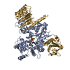
Mass: 507.181 Da / Num. of mol.: 1 / Source method: obtained synthetically / Formula: C10H16N5O13P3 / Comment: ATP, energy-carrying molecule*YM
#4: Chemical ChemComp-CA / CALCIUM ION


Mass: 40.078 Da / Num. of mol.: 3 / Source method: obtained synthetically / Formula: Ca
#5: Water ChemComp-HOH / water / Water


Mass: 18.015 Da / Num. of mol.: 208 / Source method: isolated from a natural source / Formula: H2O

-
Experimental details

-
Experiment

ExperimentMethod: X-RAY DIFFRACTION

-
Sample preparation

CrystalDensity Matthews: 3.07 Å3/Da / Density % sol: 59.98 %
Crystal growTemperature: 289 K / Method: vapor diffusion, hanging drop / pH: 7.5 / Details: 0.25 M sodium chloride, 12% w/v PEG3350 / PH range: 7.5

-
Data collection

DiffractionMean temperature: 100 K
Diffraction sourceSource: SYNCHROTRON / Site: NSLS / Beamline: X6A / Wavelength: 1.0781 Å
DetectorType: ADSC QUANTUM 270 / Detector: CCD / Date: Jan 22, 2013
RadiationMonochromator: channel cut Si(111) / Protocol: SINGLE WAVELENGTH / Monochromatic (M) / Laue (L): M / Scattering type: x-ray
Radiation wavelengthWavelength: 1.0781 Å / Relative weight: 1
ReflectionResolution: 2.3→45 Å / Num. obs: 43679 / % possible obs: 99.9 % / Redundancy: 12.8 % / Rmerge(I) obs: 0.063 / Net I/σ(I): 32.8
Reflection shellResolution: 2.3→2.44 Å / Redundancy: 12.2 % / Rmerge(I) obs: 0.57 / Mean I/σ(I) obs: 3.1 / % possible all: 100

-
Processing

Software
NameVersionClassification
PHENIX(phenix.refine: 1.8.4_1496)refinement
HKL-2000data scaling
Cootmodel building
RefinementMethod to determine structure: MOLECULAR REPLACEMENT
Starting model: PDB entry 1IO0
Resolution: 2.3→45 Å / SU ML: 0.18 / Cross valid method: FREE R-VALUE / σ(F): 1.34 / Phase error: 21.01 / Stereochemistry target values: ML
RfactorNum. reflection% reflection
Rfree0.2005 1998 4.58 %
Rwork0.1682 --
obs0.1697 43647 99.7 %
Solvent computationShrinkage radii: 0.9 Å / VDW probe radii: 1.11 Å / Solvent model: FLAT BULK SOLVENT MODEL
Refinement stepCycle: LAST / Resolution: 2.3→45 Å
ProteinNucleic acidLigandSolventTotal
Num. atoms5325 0 34 208 5567
Refine LS restraints
Refine-IDTypeDev idealNumber
X-RAY DIFFRACTIONf_bond_d0.0145527
X-RAY DIFFRACTIONf_angle_d1.4317504
X-RAY DIFFRACTIONf_dihedral_angle_d17.1782091
X-RAY DIFFRACTIONf_chiral_restr0.055831
X-RAY DIFFRACTIONf_plane_restr0.008971
LS refinement shell
Resolution (Å)Rfactor RfreeNum. reflection RfreeRfactor RworkNum. reflection RworkRefine-ID% reflection obs (%)
2.2982-2.35570.27671360.252836X-RAY DIFFRACTION97
2.3557-2.41940.28961410.23052936X-RAY DIFFRACTION100
2.4194-2.49060.23691410.21232944X-RAY DIFFRACTION100
2.4906-2.5710.23431410.19472921X-RAY DIFFRACTION100
2.571-2.66280.23311400.19122962X-RAY DIFFRACTION100
2.6628-2.76940.25541410.19812934X-RAY DIFFRACTION100
2.7694-2.89550.23331430.19032978X-RAY DIFFRACTION100
2.8955-3.04810.23331430.20462961X-RAY DIFFRACTION100
3.0481-3.2390.24371420.19732961X-RAY DIFFRACTION100
3.239-3.4890.19711420.18572968X-RAY DIFFRACTION100
3.489-3.83990.19641450.16863010X-RAY DIFFRACTION100
3.8399-4.39520.17791430.13262999X-RAY DIFFRACTION100
4.3952-5.53580.17351470.12893041X-RAY DIFFRACTION100
5.5358-44.88480.14961530.14493198X-RAY DIFFRACTION100
Refinement TLS params.

Method: refined / Refine-ID: X-RAY DIFFRACTION

IDL112)L122)L132)L222)L232)L332)S11 (Å °)S12 (Å °)S13 (Å °)S21 (Å °)S22 (Å °)S23 (Å °)S31 (Å °)S32 (Å °)S33 (Å °)T112)T122)T132)T222)T232)T332)Origin x (Å)Origin y (Å)Origin z (Å)
10.77310.38620.46771.73030.18950.98320.09260.0976-0.3263-0.2351-0.0926-0.05350.30380.0331-0.00430.37510.06610.04310.3413-0.0650.4501-1.4431-9.9781-18.0412
21.4090.0934-0.44761.6616-0.04060.7330.11490.0467-0.1103-0.4211-0.19130.21650.094-0.01980.10410.3140.03840.00970.2928-0.03360.274-6.19536.3213-16.7662
32.2276-0.3972-0.51591.8933-0.20262.45630.23910.11390.0878-1.0162-0.1619-0.38920.07750.3403-0.13630.64550.05630.2420.38650.02810.32318.941614.0027-28.9172
40.5547-0.03540.23271.89640.39981.15360.1393-0.09610.02890.0634-0.15810.1256-0.0674-0.0194-0.00670.2450.00290.07180.3333-0.03650.3508-8.12488.4194-6.291
52.6217-1.09680.68132.75230.41890.80040.3557-1.114-0.41070.4807-0.45040.27530.4039-0.72580.44060.4228-0.13220.13980.9225-0.23520.2956-10.006619.753311.0369
61.17791.27441.98111.66182.46524.25790.5871-0.6641-0.09790.5221-0.455-0.54490.2574-0.6150.1170.4476-0.14710.02410.616-0.06190.3170.518917.240713.6815
71.7306-0.29560.20461.35490.16261.7598-0.0734-0.35480.59060.3749-0.2067-0.05990.1187-0.4670.03060.1948-0.06380.05150.5475-0.20010.4296-3.123826.9086.7245
83.6426-1.3591-0.06062.2138-0.04531.10590.0934-0.47250.05070.0302-0.03990.08180.1285-0.26170.08610.2297-0.05270.060.4049-0.06870.3061-5.320616.44331.6451
91.13160.33720.82881.55420.09840.58540.0057-0.2290.86930.0036-0.16090.166-0.0699-0.06430.09230.22420.01080.0210.3948-0.12870.451-2.541229.38121.2579
101.82660.76591.02351.6126-0.59861.34560.1237-0.39490.12650.5112-0.2606-0.04760.10630.00710.07450.3034-0.0433-0.00250.5067-0.08360.39198.424623.00699.1759
110.1043-0.0226-0.01670.01890.0241-0.0297-0.3565-0.31070.0639-0.62330.1568-1.40970.08670.71-0.14670.78760.22840.04960.8741-0.01561.13954.7006-26.5804-21.4312
121.62970.3717-0.28492.7559-0.45182.7442-0.1565-0.0487-0.3326-0.25970.04240.04230.82650.120.09080.8410.04670.01830.3255-0.03710.4512-14.0159-35.2293-23.8015
131.2793-0.347-0.79231.19280.42.21760.13010.20250.1866-0.58180.01530.37820.0948-0.2532-0.17080.67020.0207-0.08030.4068-0.00250.5478-21.2443-19.5565-28.4206
143.09-0.86291.13083.98710.90471.74170.4412-0.15250.1734-0.1369-0.19760.46780.064-0.0841-0.07180.51990.01360.06190.4169-0.07120.7639-25.8803-9.067-13.9243
Refinement TLS group
IDRefine-IDRefine TLS-IDSelection details
1X-RAY DIFFRACTION1chain 'A' and (resid 5 through 97 )
2X-RAY DIFFRACTION2chain 'A' and (resid 98 through 196 )
3X-RAY DIFFRACTION3chain 'A' and (resid 197 through 320 )
4X-RAY DIFFRACTION4chain 'A' and (resid 321 through 375 )
5X-RAY DIFFRACTION5chain 'G' and (resid 52 through 73 )
6X-RAY DIFFRACTION6chain 'G' and (resid 74 through 85 )
7X-RAY DIFFRACTION7chain 'G' and (resid 86 through 121 )
8X-RAY DIFFRACTION8chain 'G' and (resid 122 through 138 )
9X-RAY DIFFRACTION9chain 'G' and (resid 139 through 160 )
10X-RAY DIFFRACTION10chain 'G' and (resid 161 through 177 )
11X-RAY DIFFRACTION11chain 'G' and (resid 1170 through 1182 )
12X-RAY DIFFRACTION12chain 'G' and (resid 1183 through 1237 )
13X-RAY DIFFRACTION13chain 'G' and (resid 1238 through 1321 )
14X-RAY DIFFRACTION14chain 'G' and (resid 1322 through 1349 )

+
About Yorodumi

-
News

-
Feb 9, 2022. New format data for meta-information of EMDB entries

New format data for meta-information of EMDB entries

  • Version 3 of the EMDB header file is now the official format.
  • The previous official version 1.9 will be removed from the archive.

Related info.:EMDB header

External links:wwPDB to switch to version 3 of the EMDB data model

-
Aug 12, 2020. Covid-19 info

Covid-19 info

URL: https://pdbj.org/emnavi/covid19.php

New page: Covid-19 featured information page in EM Navigator.

Related info.:Covid-19 info / Mar 5, 2020. Novel coronavirus structure data

+
Mar 5, 2020. Novel coronavirus structure data

Novel coronavirus structure data

Related info.:Yorodumi Speices / Aug 12, 2020. Covid-19 info

External links:COVID-19 featured content - PDBj / Molecule of the Month (242):Coronavirus Proteases

+
Jan 31, 2019. EMDB accession codes are about to change! (news from PDBe EMDB page)

EMDB accession codes are about to change! (news from PDBe EMDB page)

  • The allocation of 4 digits for EMDB accession codes will soon come to an end. Whilst these codes will remain in use, new EMDB accession codes will include an additional digit and will expand incrementally as the available range of codes is exhausted. The current 4-digit format prefixed with “EMD-” (i.e. EMD-XXXX) will advance to a 5-digit format (i.e. EMD-XXXXX), and so on. It is currently estimated that the 4-digit codes will be depleted around Spring 2019, at which point the 5-digit format will come into force.
  • The EM Navigator/Yorodumi systems omit the EMD- prefix.

Related info.:Q: What is EMD? / ID/Accession-code notation in Yorodumi/EM Navigator

External links:EMDB Accession Codes are Changing Soon! / Contact to PDBj

+
Jul 12, 2017. Major update of PDB

Major update of PDB

  • wwPDB released updated PDB data conforming to the new PDBx/mmCIF dictionary.
  • This is a major update changing the version number from 4 to 5, and with Remediation, in which all the entries are updated.
  • In this update, many items about electron microscopy experimental information are reorganized (e.g. em_software).
  • Now, EM Navigator and Yorodumi are based on the updated data.

External links:wwPDB Remediation / Enriched Model Files Conforming to OneDep Data Standards Now Available in the PDB FTP Archive

-
Yorodumi

Thousand views of thousand structures

  • Yorodumi is a browser for structure data from EMDB, PDB, SASBDB, etc.
  • This page is also the successor to EM Navigator detail page, and also detail information page/front-end page for Omokage search.
  • The word "yorodu" (or yorozu) is an old Japanese word meaning "ten thousand". "mi" (miru) is to see.

Related info.:EMDB / PDB / SASBDB / Comparison of 3 databanks / Yorodumi Search / Aug 31, 2016. New EM Navigator & Yorodumi / Yorodumi Papers / Jmol/JSmol / Function and homology information / Changes in new EM Navigator and Yorodumi

Read more