 Movie
Movie Controller

[English] 日本語
 Yorodumi
- PDB-4en9: Crystal structure of HA70 (HA3) subcomponent of Clostridium botul... -
+ Open data

- Basic information
| Entry | Database: PDB / ID: 4en9 | |||||||||
|---|---|---|---|---|---|---|---|---|---|---|
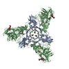
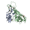
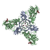
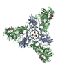
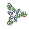
| Title | Crystal structure of HA70 (HA3) subcomponent of Clostridium botulinum type C progenitor toxin in complex with alpha 2-6-sialyllactosamine | |||||||||
 Components Components | (Hemagglutinin components HA-22/23/53) x 2 | |||||||||
 Keywords Keywords | SUGAR BINDING PROTEIN / Carbohydrate/Sugar Binding | |||||||||
| Function / homology |  Function and homology information Function and homology informationsymbiont-mediated migration across host transepithelium / extracellular region Similarity search - Function | |||||||||
| Biological species |   Clostridium botulinum (bacteria) Clostridium botulinum (bacteria) | |||||||||
| Method |  X-RAY DIFFRACTION / SYNCHROTRON / MOLECULAR REPLACEMENT / Resolution: 2.64 Å X-RAY DIFFRACTION / SYNCHROTRON / MOLECULAR REPLACEMENT / Resolution: 2.64 Å | |||||||||
 Authors Authors | Yamashita, S. / Yoshida, H. / Tonozuka, T. / Nishikawa, A. / Kamitori, S. | |||||||||
 Citation Citation | Journal: Febs Lett. / Year: 2012 Title: Carbohydrate recognition mechanism of HA70 from Clostridium botulinum deduced from X-ray structures in complexes with sialylated oligosaccharides Authors: Yamashita, S. / Yoshida, H. / Uchiyama, N. / Nakakita, Y. / Nakakita, S. / Tonozuka, T. / Oguma, K. / Nishikawa, A. / Kamitori, S. #1: Journal: J.Mol.Biol. / Year: 2009Title: Crystal structure of the HA3 subcomponent of Clostridium botulinum type C progenitor toxin Authors: Nakamura, T. / Kotani, M. / Tonozuka, T. / Ide, A. / Oguma, K. / Nishikawa, A. | |||||||||
| History |
|
- Structure visualization
| Structure viewer | Molecule: MolmilJmol/JSmol |
|---|
- Downloads & links
-Download
| PDBx/mmCIF format | 4en9.cif.gz | 143.9 KB | Display | PDBx/mmCIF format |
|---|---|---|---|---|
| PDB format | pdb4en9.ent.gz | 108.7 KB | Display | PDB format |
| PDBx/mmJSON format | 4en9.json.gz | Tree view | PDBx/mmJSON format | |
| Others |  Other downloads Other downloads |
-Validation report
| Arichive directory | https://data.pdbj.org/pub/pdb/validation_reports/en/4en9ftp://data.pdbj.org/pub/pdb/validation_reports/en/4en9 | HTTPS FTP |
|---|
-Related structure data
| Related structure data |  4en6C  4en7C  4en8C  2zs6S S: Starting model for refinement C: citing same article ( |
|---|---|
| Similar structure data |


-Links
 PDBj
PDBj- Assembly
| Deposited unit | 
| ||||||||
|---|---|---|---|---|---|---|---|---|---|
| 1 | 
| ||||||||
| Unit cell |
|
-Components
| #1: Protein | Mass: 26050.873 Da / Num. of mol.: 1 / Fragment: HA22-23(HA3a) Source method: isolated from a genetically manipulated source Source: (gene. exp.) Clostridium botulinum (bacteria) / Gene: HA-70 / Plasmid: pMAL-cRI / Production host: | ||
|---|---|---|---|
| #2: Protein | Mass: 47333.520 Da / Num. of mol.: 1 / Fragment: HA53(HA3b) Source method: isolated from a genetically manipulated source Source: (gene. exp.) Clostridium botulinum (bacteria) / Gene: HA-70 / Plasmid: pMAL-cRI / Production host: | ||
| #3: Polysaccharide | N-acetyl-alpha-neuraminic acid-(2-6)-beta-D-galactopyranose-(1-4)-2-acetamido-2-deoxy-beta-D-glucopyranose / 6'-sialyl-N-acetyllactosamine  Source method: isolated from a genetically manipulated source Details: oligosaccharide / References: 6'-sialyl-N-acetyllactosamine | ||
| #4: Chemical | ChemComp-MRD / (   Mass: 118.174 Da / Num. of mol.: 5 / Source method: obtained synthetically / Formula: C6H14O2 / Comment: precipitant*YM Mass: 118.174 Da / Num. of mol.: 5 / Source method: obtained synthetically / Formula: C6H14O2 / Comment: precipitant*YM#5: Water | ChemComp-HOH / |  Mass: 18.015 Da / Num. of mol.: 285 / Source method: isolated from a natural source / Formula: H2O Mass: 18.015 Da / Num. of mol.: 285 / Source method: isolated from a natural source / Formula: H2O |
-Experimental details
-Experiment
| Experiment | Method: X-RAY DIFFRACTION / Number of used crystals: 1 |
|---|
- Sample preparation
| Crystal | Density Matthews: 4.91 Å3/Da / Density % sol: 74.96 % |
|---|---|
| Crystal grow | Temperature: 293 K / Method: vapor diffusion, hanging drop / pH: 4.6 Details: 12%(v/v) 2-methyl-2,4-pentendiol, 20mM CaCl2, 100mM sodium acetate, pH 4.6, VAPOR DIFFUSION, HANGING DROP, temperature 293K |
-Data collection
| Diffraction | Mean temperature: 100 K |
|---|---|
| Diffraction source | Source: SYNCHROTRON / Site: Photon Factory  / Beamline: AR-NE3A / Wavelength: 1 Å / Beamline: AR-NE3A / Wavelength: 1 Å |
| Detector | Type: ADSC QUANTUM 270 / Detector: CCD / Date: Jun 17, 2011 |
| Radiation | Protocol: SINGLE WAVELENGTH / Monochromatic (M) / Laue (L): M / Scattering type: x-ray |
| Radiation wavelength | Wavelength: 1 Å / Relative weight: 1 |
| Reflection | Resolution: 2.64→50 Å / Num. all: 42102 / Num. obs: 42102 / % possible obs: 99.7 % / Observed criterion σ(F): 0 / Observed criterion σ(I): 0 / Redundancy: 10.7 % / Biso Wilson estimate: 45.3 Å2 / Rmerge(I) obs: 0.075 / Net I/σ(I): 23.7 |
| Reflection shell | Resolution: 2.64→2.69 Å / Redundancy: 10.3 % / Rmerge(I) obs: 0.372 / Mean I/σ(I) obs: 7.9 / Num. unique all: 2132 / % possible all: 100 |
- Processing
| Software |
| |||||||||||||||||||||||||
|---|---|---|---|---|---|---|---|---|---|---|---|---|---|---|---|---|---|---|---|---|---|---|---|---|---|---|
| Refinement | Method to determine structure: MOLECULAR REPLACEMENT Starting model: PDB ENTRY 2ZS6 Resolution: 2.64→33.26 Å / Rfactor Rfree error: 0.003 / Data cutoff high absF: 88421.32 / Data cutoff low absF: 0 / Isotropic thermal model: RESTRAINED / Cross valid method: THROUGHOUT / σ(F): 0 / σ(I): 0 / Stereochemistry target values: Engh & Huber
| |||||||||||||||||||||||||
| Solvent computation | Solvent model: FLAT MODEL / Bsol: 43.4246 Å2 / ksol: 0.320009 e/Å3 | |||||||||||||||||||||||||
| Displacement parameters | Biso mean: 52 Å2
| |||||||||||||||||||||||||
| Refine analyze |
| |||||||||||||||||||||||||
| Refinement step | Cycle: LAST / Resolution: 2.64→33.26 Å
| |||||||||||||||||||||||||
| Refine LS restraints |
| |||||||||||||||||||||||||
| LS refinement shell | Resolution: 2.64→2.81 Å / Rfactor Rfree error: 0.012 / Total num. of bins used: 6
| |||||||||||||||||||||||||
| Xplor file |
|
