 Movie
Movie Controller


+ Open data

- Basic information
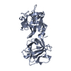
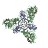
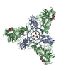
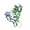
| Entry | Database: PDB / ID: 2zs6 | |||||||||
|---|---|---|---|---|---|---|---|---|---|---|
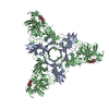
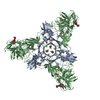
| Title | HA3 subcomponent of botulinum type C progenitor toxin | |||||||||
 Components Components | (Hemagglutinin components HA3) x 2 | |||||||||
 Keywords Keywords | TOXIN / LECTIN / HEMAGGLUTININ / BOTULINUM TOXIN | |||||||||
| Function / homology |  Function and homology information Function and homology informationsymbiont-mediated migration across host transepithelium / extracellular region Similarity search - Function | |||||||||
| Biological species |   Clostridium botulinum (bacteria) Clostridium botulinum (bacteria) | |||||||||
| Method |  X-RAY DIFFRACTION / SYNCHROTRON / MOLECULAR REPLACEMENT / Resolution: 2.6 Å X-RAY DIFFRACTION / SYNCHROTRON / MOLECULAR REPLACEMENT / Resolution: 2.6 Å | |||||||||
 Authors Authors | Nakamura, T. / Tonozuka, T. / Kotani, M. / Oguma, K. / Nishikawa, A. | |||||||||
 Citation Citation | Journal: J.Mol.Biol. / Year: 2009 Title: Crystal Structure of the HA3 Subcomponent of Clostridium botulinum Type C Progenitor Toxin Authors: Nakamura, T. / Kotani, M. / Tonozuka, T. / Ide, A. / Oguma, K. / Nishikawa, A. #1: Journal: J.Mol.Biol. / Year: 2008Title: Sugar-binding sites of the HA1 subcomponent of Clostridium botulinum type C progenitor toxin. Authors: Nakamura, T. / Tonozuka, T. / Ide, A. / Yuzawa, T. / Oguma, K. / Nishikawa, A. #2: Journal: Biochim.Biophys.Acta / Year: 2007 Title: Binding properties of Clostridium botulinum type C progenitor toxin to mucins. Authors: Nakamura, T. / Takada, N. / Tonozuka, T. / Sakano, Y. / Oguma, K. / Nishikawa, A. | |||||||||
| History |
|
- Structure visualization
| Structure viewer | Molecule: MolmilJmol/JSmol |
|---|
- Downloads & links
-Download
| PDBx/mmCIF format | 2zs6.cif.gz | 137 KB | Display | PDBx/mmCIF format |
|---|---|---|---|---|
| PDB format | pdb2zs6.ent.gz | 105.8 KB | Display | PDB format |
| PDBx/mmJSON format | 2zs6.json.gz | Tree view | PDBx/mmJSON format | |
| Others |  Other downloads Other downloads |
-Validation report
| Arichive directory | https://data.pdbj.org/pub/pdb/validation_reports/zs/2zs6ftp://data.pdbj.org/pub/pdb/validation_reports/zs/2zs6 | HTTPS FTP |
|---|
-Related structure data


-Links
 PDBj
PDBj
- Assembly
| Deposited unit | 
| ||||||||
|---|---|---|---|---|---|---|---|---|---|
| 1 | 
| ||||||||
| Unit cell |
|
-Components
| #1: Protein | Mass: 23955.641 Da / Num. of mol.: 1 / Fragment: HA3a Source method: isolated from a genetically manipulated source Source: (gene. exp.) Clostridium botulinum (bacteria) / Strain: type C / Plasmid: pMAL-cRI / Production host: |
|---|---|
| #2: Protein | Mass: 47333.520 Da / Num. of mol.: 1 / Fragment: HA3b Source method: isolated from a genetically manipulated source Source: (gene. exp.) Clostridium botulinum (bacteria) / Strain: type C / Plasmid: pMAL-cRI / Production host: |
| #3: Water | ChemComp-HOH /  Mass: 18.015 Da / Num. of mol.: 228 / Source method: isolated from a natural source / Formula: H2O Mass: 18.015 Da / Num. of mol.: 228 / Source method: isolated from a natural source / Formula: H2O |
-Experimental details
-Experiment
| Experiment | Method: X-RAY DIFFRACTION / Number of used crystals: 1 |
|---|
- Sample preparation
| Crystal | Density Matthews: 5.2961 Å3/Da / Density % sol: 76.77536 % |
|---|---|
| Crystal grow | Temperature: 293 K / Method: vapor diffusion, hanging drop / pH: 4.4 Details: 1.2M AMMONIUM PHOSPHATE, 0.1M SODIUM CITRATE, pH 4.4, VAPOR DIFFUSION, HANGING DROP, temperature 293K |
-Data collection
| Diffraction | Mean temperature: 100 K |
|---|---|
| Diffraction source | Source: SYNCHROTRON / Site: Photon Factory  / Beamline: BL-5A / Wavelength: 1 Å / Beamline: BL-5A / Wavelength: 1 Å |
| Detector | Type: ADSC QUANTUM 4 / Detector: CCD / Date: Jun 2, 2006 |
| Radiation | Protocol: SINGLE WAVELENGTH / Monochromatic (M) / Laue (L): M / Scattering type: x-ray |
| Radiation wavelength | Wavelength: 1 Å / Relative weight: 1 |
| Reflection | Resolution: 2.6→50 Å / Num. obs: 44256 / % possible obs: 100 % / Redundancy: 50.9 % / Biso Wilson estimate: 51.6 Å2 / Rsym value: 0.057 |
| Reflection shell | Resolution: 2.6→2.69 Å / Mean I/σ(I) obs: 5.9 / Rsym value: 0.311 / % possible all: 100 |
- Processing
| Software |
| ||||||||||||||||||||||||||||||||||||||||||||||||||||||||||||||||||||||||||||||||
|---|---|---|---|---|---|---|---|---|---|---|---|---|---|---|---|---|---|---|---|---|---|---|---|---|---|---|---|---|---|---|---|---|---|---|---|---|---|---|---|---|---|---|---|---|---|---|---|---|---|---|---|---|---|---|---|---|---|---|---|---|---|---|---|---|---|---|---|---|---|---|---|---|---|---|---|---|---|---|---|---|---|
| Refinement | Method to determine structure: MOLECULAR REPLACEMENT Starting model: A ROUGH MODEL BUILED BY MAD DATA SET Resolution: 2.6→47 Å / Rfactor Rfree error: 0.004 / Data cutoff high absF: 2346465.12 / Data cutoff low absF: 0 / Isotropic thermal model: RESTRAINED / Cross valid method: THROUGHOUT / σ(F): 0 / Stereochemistry target values: Engh & Huber / Details: BULK SOLVENT MODEL USED
| ||||||||||||||||||||||||||||||||||||||||||||||||||||||||||||||||||||||||||||||||
| Solvent computation | Solvent model: FLAT MODEL / Bsol: 43.7214 Å2 / ksol: 0.35 e/Å3 | ||||||||||||||||||||||||||||||||||||||||||||||||||||||||||||||||||||||||||||||||
| Displacement parameters | Biso mean: 57.6 Å2
| ||||||||||||||||||||||||||||||||||||||||||||||||||||||||||||||||||||||||||||||||
| Refine analyze |
| ||||||||||||||||||||||||||||||||||||||||||||||||||||||||||||||||||||||||||||||||
| Refinement step | Cycle: LAST / Resolution: 2.6→47 Å
| ||||||||||||||||||||||||||||||||||||||||||||||||||||||||||||||||||||||||||||||||
| Refine LS restraints |
| ||||||||||||||||||||||||||||||||||||||||||||||||||||||||||||||||||||||||||||||||
| LS refinement shell | Resolution: 2.6→2.76 Å / Rfactor Rfree error: 0.011 / Total num. of bins used: 6
| ||||||||||||||||||||||||||||||||||||||||||||||||||||||||||||||||||||||||||||||||
| Xplor file |
|
