| 登録情報 | データベース: PDB / ID: 4z94
|
|---|
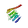
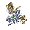
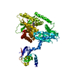
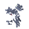
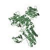
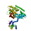
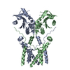
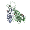
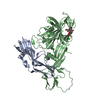
| タイトル | Actin Complex With a Chimera of Tropomodulin-1 and Leiomodin-1 Actin-Binding Site 2 |
|---|
 要素 要素 | - Actin, alpha skeletal muscle
 - Gelsolin, Tropomodulin-1, Leiomodin-1 chimera
|
|---|
 キーワード キーワード | PROTEIN BINDING/STRUCTURAL PROTEIN / Actin Nucleator / ATP-actin / PROTEIN BINDING-STRUCTURAL PROTEIN complex |
|---|
| 機能・相同性 |  機能・相同性情報 機能・相同性情報
pointed-end actin filament capping / lens fiber cell development / myofibril assembly / actin nucleation / striated muscle atrophy / regulation of establishment of T cell polarity / regulation of plasma membrane raft polarization / regulation of receptor clustering / renal protein absorption / positive regulation of keratinocyte apoptotic process ...pointed-end actin filament capping / lens fiber cell development / myofibril assembly / actin nucleation / striated muscle atrophy / regulation of establishment of T cell polarity / regulation of plasma membrane raft polarization / regulation of receptor clustering / renal protein absorption / positive regulation of keratinocyte apoptotic process / positive regulation of protein processing in phagocytic vesicle / positive regulation of actin nucleation / phosphatidylinositol 3-kinase catalytic subunit binding / positive regulation of cysteine-type endopeptidase activity involved in apoptotic signaling pathway / actin cap / sequestering of actin monomers / regulation of podosome assembly / myosin II binding / negative regulation of viral entry into host cell / actin filament severing / actin filament capping / barbed-end actin filament capping / actin filament depolymerization / actin polymerization or depolymerization / cell projection assembly / cardiac muscle cell contraction / Striated Muscle Contraction / podosome / Sensory processing of sound by outer hair cells of the cochlea / relaxation of cardiac muscle / cytoskeletal motor activator activity / phagocytosis, engulfment / cortical actin cytoskeleton / tropomyosin binding / cortical cytoskeleton / myofibril / myosin heavy chain binding / positive regulation of actin filament polymerization / mesenchyme migration / troponin I binding / hepatocyte apoptotic process / actin filament bundle / filamentous actin / actin filament bundle assembly / skeletal muscle thin filament assembly / striated muscle thin filament / skeletal muscle myofibril / cilium assembly / actin monomer binding / 筋形質 / Smooth Muscle Contraction / Caspase-mediated cleavage of cytoskeletal proteins / skeletal muscle fiber development / phagocytic vesicle / stress fiber / titin binding / phosphatidylinositol-4,5-bisphosphate binding / response to muscle stretch / actin filament polymerization / sarcomere / adult locomotory behavior / filopodium / muscle contraction / central nervous system development / actin filament organization / マイクロフィラメント / 加水分解酵素; 酸無水物に作用; 酸無水物に作用・細胞または細胞小器官の運動に関与 / protein destabilization / cellular response to type II interferon / calcium-dependent protein binding / actin filament binding / マイクロフィラメント / lamellipodium / cell body / actin binding / blood microparticle / secretory granule lumen / ficolin-1-rich granule lumen / amyloid fibril formation / 細胞骨格 / hydrolase activity / Amyloid fiber formation / protein domain specific binding / focal adhesion / calcium ion binding / Neutrophil degranulation / positive regulation of gene expression / magnesium ion binding / extracellular space / extracellular exosome / extracellular region / ATP binding / 生体膜 / identical protein binding / 細胞膜 / 細胞質基質 / 細胞質類似検索 - 分子機能 Tropomodulin / Tropomodulin / WH2 motif / Wiskott Aldrich syndrome homology region 2 / WH2 domain / WH2 domain profile. / Actin; Chain A, domain 2 / Actin; Chain A, domain 2 / Villin/Gelsolin / Gelsolin homology domain ...Tropomodulin / Tropomodulin / WH2 motif / Wiskott Aldrich syndrome homology region 2 / WH2 domain / WH2 domain profile. / Actin; Chain A, domain 2 / Actin; Chain A, domain 2 / Villin/Gelsolin / Gelsolin homology domain / Severin / Severin / Gelsolin-like domain / Gelsolin repeat / ADF-H/Gelsolin-like domain superfamily / Leucine-rich repeat, LRR (right-handed beta-alpha superhelix) / リボヌクレアーゼインヒビター / ATPase, substrate binding domain, subdomain 4 / Actin; Chain A, domain 4 / Alpha-Beta Horseshoe / ATPase, nucleotide binding domain / Actins signature 1. / Actin, conserved site / Actins signature 2. / Actin/actin-like conserved site / Actins and actin-related proteins signature. / アクチン / Actin family / アクチン / Leucine-rich repeat domain superfamily / ATPase, nucleotide binding domain / Nucleotidyltransferase; domain 5 / Roll / Alpha-Beta Complex / 2-Layer Sandwich / 3-Layer(aba) Sandwich / Mainly Beta / Alpha Beta類似検索 - ドメイン・相同性 ADENOSINE-5'-TRIPHOSPHATE / Gelsolin / Tropomodulin-1 / Leiomodin-1 / Actin, alpha skeletal muscle類似検索 - 構成要素 |
|---|
| 生物種 |  Homo sapiens (ヒト) Homo sapiens (ヒト)
  Oryctolagus cuniculus (ウサギ) Oryctolagus cuniculus (ウサギ) |
|---|
| 手法 | X線回折 / 分子置換 / 解像度: 2.4 Å |
|---|
 データ登録者 データ登録者 | Rebowski, G. / Boczkowska, M. / Dominguez, R. |
|---|
| 資金援助 |  米国, 1件 米国, 1件 | 組織 | 認可番号 | 国 |
|---|
| National Institutes of Health/National Institute of General Medical Sciences (NIH/NIGMS) | R01 GM073791 | 米国 |
|
|---|
 引用 引用 | ジャーナル: Nat Commun / 年: 2015
タイトル: How Leiomodin and Tropomodulin use a common fold for different actin assembly functions.
著者: Boczkowska, M. / Rebowski, G. / Kremneva, E. / Lappalainen, P. / Dominguez, R. |
|---|
| 履歴 | | 登録 | 2015年4月9日 | 登録サイト: RCSB / 処理サイト: RCSB |
|---|
| 改定 1.0 | 2015年10月21日 | Provider: repository / タイプ: Initial release |
|---|
| 改定 1.1 | 2017年9月6日 | Group: Author supporting evidence / Derived calculations / カテゴリ: pdbx_audit_support / pdbx_struct_oper_list
Item: _pdbx_audit_support.funding_organization / _pdbx_struct_oper_list.symmetry_operation |
|---|
| 改定 1.2 | 2018年3月7日 | Group: Data collection / カテゴリ: diffrn_source / Item: _diffrn_source.source |
|---|
| 改定 1.3 | 2019年12月25日 | Group: Author supporting evidence / カテゴリ: pdbx_audit_support / Item: _pdbx_audit_support.funding_organization |
|---|
| 改定 1.4 | 2023年9月27日 | Group: Data collection / Database references ...Data collection / Database references / Derived calculations / Refinement description
カテゴリ: chem_comp_atom / chem_comp_bond ...chem_comp_atom / chem_comp_bond / database_2 / pdbx_initial_refinement_model / pdbx_struct_conn_angle / struct_conn
Item: _database_2.pdbx_DOI / _database_2.pdbx_database_accession ..._database_2.pdbx_DOI / _database_2.pdbx_database_accession / _pdbx_struct_conn_angle.ptnr1_auth_asym_id / _pdbx_struct_conn_angle.ptnr1_auth_comp_id / _pdbx_struct_conn_angle.ptnr1_auth_seq_id / _pdbx_struct_conn_angle.ptnr1_label_asym_id / _pdbx_struct_conn_angle.ptnr1_label_atom_id / _pdbx_struct_conn_angle.ptnr1_label_comp_id / _pdbx_struct_conn_angle.ptnr1_label_seq_id / _pdbx_struct_conn_angle.ptnr2_auth_asym_id / _pdbx_struct_conn_angle.ptnr2_auth_seq_id / _pdbx_struct_conn_angle.ptnr2_label_asym_id / _pdbx_struct_conn_angle.ptnr3_auth_asym_id / _pdbx_struct_conn_angle.ptnr3_auth_comp_id / _pdbx_struct_conn_angle.ptnr3_auth_seq_id / _pdbx_struct_conn_angle.ptnr3_label_asym_id / _pdbx_struct_conn_angle.ptnr3_label_atom_id / _pdbx_struct_conn_angle.ptnr3_label_comp_id / _pdbx_struct_conn_angle.ptnr3_label_seq_id / _pdbx_struct_conn_angle.value / _struct_conn.pdbx_dist_value / _struct_conn.ptnr1_auth_asym_id / _struct_conn.ptnr1_auth_comp_id / _struct_conn.ptnr1_auth_seq_id / _struct_conn.ptnr1_label_asym_id / _struct_conn.ptnr1_label_atom_id / _struct_conn.ptnr1_label_comp_id / _struct_conn.ptnr1_label_seq_id / _struct_conn.ptnr2_auth_asym_id / _struct_conn.ptnr2_auth_comp_id / _struct_conn.ptnr2_auth_seq_id / _struct_conn.ptnr2_label_asym_id / _struct_conn.ptnr2_label_atom_id / _struct_conn.ptnr2_label_comp_id / _struct_conn.ptnr2_label_seq_id |
|---|
|
|---|
 ムービー
ムービー コントローラー


 データを開く
 基本情報
基本情報 構造の表示
構造の表示 ダウンロードとリンク
 その他のダウンロード
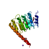


 PDBj
PDBj


 集合体
集合体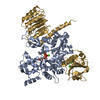


 分子量: 507.181 Da / 分子数: 1 / 由来タイプ: 天然 / 式: C10H16N5O13P3 / コメント: ATP, エネルギー貯蔵分子*YM
 分子量: 40.078 Da / 分子数: 3 / 由来タイプ: 合成 / 式: Ca
 分子量: 18.015 Da / 分子数: 182 / 由来タイプ: 天然 / 式: H2O
 試料調製
試料調製 解析
解析