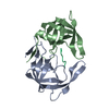
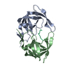
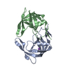
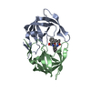
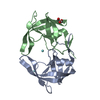
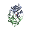
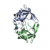
Entry Database : PDB / ID : 4z4xTitle Crystal Structure of Multidrug Resistant HIV-1 Protease Clinical Isolate PR20D25N with Open Flap Protease Keywords / Function / homology Function Domain/homology Component
/ / / / / / / / / / / / / / / / / / / / Biological species Method / / / Resolution : 1.75 Å Authors Chang, Y.C. / Shen, C.-H. / Weber, I.T. Funding support Organization Grant number Country National Institutes of Health/National Institute of General Medical Sciences (NIH/NIGMS) U01GM062920
Journal : J.Mol.Graph.Model. / Year : 2015Title : Conformational variation of an extreme drug resistant mutant of HIV protease.Authors : Shen, C.H. / Chang, Y.C. / Agniswamy, J. / Harrison, R.W. / Weber, I.T. History Deposition Apr 2, 2015 Deposition site / Processing site Revision 1.0 Oct 14, 2015 Provider / Type Revision 1.1 Sep 27, 2017 Group / Derived calculations / Category / pdbx_struct_oper_listItem / _pdbx_struct_oper_list.symmetry_operationRevision 1.2 Dec 25, 2019 Group / Category / Item Revision 1.3 Sep 27, 2023 Group / Database references / Refinement descriptionCategory chem_comp_atom / chem_comp_bond ... chem_comp_atom / chem_comp_bond / database_2 / pdbx_initial_refinement_model Item / _database_2.pdbx_database_accession
Show all Show less
 Movie
Movie Controller

 Yorodumi
 Open data
 Basic information
 Components
 Keywords
 Function and homology information
 Human immunodeficiency virus 1
 X-RAY DIFFRACTION /
  Authors
 United States, 1items
  Citation
 Structure visualization
 Downloads & links
 Other downloads
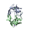


 PDBj
PDBj
 Assembly
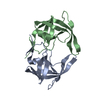

 Mass: 18.015 Da / Num. of mol.: 110 / Source method: isolated from a natural source / Formula: H2O
 Sample preparation
 Processing