 Movie
Movie Controller

[English] 日本語
 Yorodumi
- PDB-1tw7: Wide Open 1.3A Structure of a Multi-drug Resistant HIV-1 Protease... -
+ Open data

- Basic information
| Entry | Database: PDB / ID: 1tw7 | ||||||
|---|---|---|---|---|---|---|---|
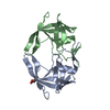
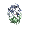
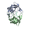
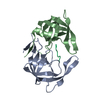
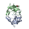
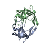
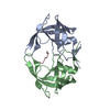
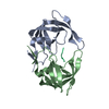
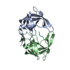
| Title | Wide Open 1.3A Structure of a Multi-drug Resistant HIV-1 Protease Represents a Novel Drug Target | ||||||
 Components Components | protease | ||||||
 Keywords Keywords | HYDROLASE / HIV protease / AIDS / polyprotein / aspartyl protease / multi-drug resistance | ||||||
| Function / homology |  Function and homology information Function and homology informationhost multivesicular body / aspartic-type endopeptidase activity / virion membrane / proteolysis Similarity search - Function | ||||||
| Biological species |   Human immunodeficiency virus 1 Human immunodeficiency virus 1 | ||||||
| Method |  X-RAY DIFFRACTION / SYNCHROTRON / MOLECULAR REPLACEMENT / Resolution: 1.3 Å X-RAY DIFFRACTION / SYNCHROTRON / MOLECULAR REPLACEMENT / Resolution: 1.3 Å | ||||||
 Authors Authors | Martin, P. / Vickrey, J.F. / Proteasa, G. / Jimenez, Y.L. / Wawrzak, Z. / Winters, M.A. / Merigan, T.C. / Kovari, L.C. | ||||||
 Citation Citation | Journal: Structure / Year: 2005 Title: Wide Open 1.3A Structure of a Multi-drug Resistant HIV-1 Protease Represents a Novel Drug Target Authors: Martin, P. / Vickrey, J.F. / Proteasa, G. / Jimenez, Y.L. / Wawrzak, Z. / Winters, M.A. / Merigan, T.C. / Kovari, L.C. #1: Journal: J.Virol. / Year: 2004Title: Crystal structures of a Multidrug-Resistant human immunodeficiency virus type 1 Protease Reveal an Expanded Active-Site Cavity Authors: Logsdon, B.C. / Vickrey, J.F. / Martin, P. / Proteasa, G. / Kopeke, J.I. / Terlecky, S.R. / Wawrzak, Z. / Winters, M.A. / Merigan, T.C. / Kovari, L.C. #2: Journal: PROTEIN EXPR.PURIF. / Year: 2003Title: HIV-1 Protease variants from 100-fold drug resistant clinical isolates: expression, purification, and crystallization. Authors: Vickrey, J.F. / Logsdon, B.C. / Proteasa, G. / Palmer, S. / Winters, M.A. / Merigan, T.C. / Kovari, L.C. | ||||||
| History |
|
- Structure visualization
| Structure viewer | Molecule: MolmilJmol/JSmol |
|---|
- Downloads & links
-Download
| PDBx/mmCIF format | 1tw7.cif.gz | 108.4 KB | Display | PDBx/mmCIF format |
|---|---|---|---|---|
| PDB format | pdb1tw7.ent.gz | 83.4 KB | Display | PDB format |
| PDBx/mmJSON format | 1tw7.json.gz | Tree view | PDBx/mmJSON format | |
| Others |  Other downloads Other downloads |
-Validation report
| Arichive directory | https://data.pdbj.org/pub/pdb/validation_reports/tw/1tw7ftp://data.pdbj.org/pub/pdb/validation_reports/tw/1tw7 | HTTPS FTP |
|---|
-Related structure data
| Related structure data |  1rq9S S: Starting model for refinement |
|---|---|
| Similar structure data |


-Links
 PDBj
PDBj- Assembly
| Deposited unit | 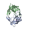
| ||||||||
|---|---|---|---|---|---|---|---|---|---|
| 1 |
| ||||||||
| Unit cell |
|
-Components
| #1: Protein | Mass: 10739.608 Da / Num. of mol.: 2 Mutation: L10I, D25N, M36V, M46L, I54V, I62V, L63P, A71V, V82A, I84V, L90M Source method: isolated from a genetically manipulated source Source: (gene. exp.) Human immunodeficiency virus 1 / Genus: Lentivirus / Gene: HIV-1 / Plasmid: pJFV769noHisKO / Species (production host): Escherichia coli / Production host:  References: GenBank: 30142908, UniProt: Q5RTL1*PLUS, HIV-1 retropepsin #2: Chemical |   Mass: 22.990 Da / Num. of mol.: 2 / Source method: obtained synthetically / Formula: Na Mass: 22.990 Da / Num. of mol.: 2 / Source method: obtained synthetically / Formula: Na#3: Water | ChemComp-HOH / |  Mass: 18.015 Da / Num. of mol.: 380 / Source method: isolated from a natural source / Formula: H2O Mass: 18.015 Da / Num. of mol.: 380 / Source method: isolated from a natural source / Formula: H2O |
|---|
-Experimental details
-Experiment
| Experiment | Method: X-RAY DIFFRACTION / Number of used crystals: 1 |
|---|
- Sample preparation
| Crystal | Density Matthews: 2.5 Å3/Da / Density % sol: 50.74 % |
|---|---|
| Crystal grow | Temperature: 295 K / Method: vapor diffusion, hanging drop / pH: 6.5 Details: sodium chloride, MES, pH 6.5, VAPOR DIFFUSION, HANGING DROP, temperature 295K |
-Data collection
| Diffraction | Mean temperature: 100 K |
|---|---|
| Diffraction source | Source: SYNCHROTRON / Site: APS  / Beamline: 17-BM / Wavelength: 1 Å / Beamline: 17-BM / Wavelength: 1 Å |
| Detector | Type: ADSC QUANTUM 210 / Detector: CCD / Date: Jul 27, 2003 |
| Radiation | Monochromator: SILCON / Protocol: SINGLE WAVELENGTH / Monochromatic (M) / Laue (L): M / Scattering type: x-ray |
| Radiation wavelength | Wavelength: 1 Å / Relative weight: 1 |
| Reflection | Resolution: 1.3→20 Å / Num. obs: 38356 / % possible obs: 74.7 % / Rsym value: 0.043 / Net I/σ(I): 30.5 |
| Reflection shell | Resolution: 1.3→1.35 Å / % possible all: 0.26 |
- Processing
| Software |
| |||||||||||||||||||||||||||||||||
|---|---|---|---|---|---|---|---|---|---|---|---|---|---|---|---|---|---|---|---|---|---|---|---|---|---|---|---|---|---|---|---|---|---|---|
| Refinement | Method to determine structure: MOLECULAR REPLACEMENT Starting model: PDB entry 1RQ9 Resolution: 1.3→20 Å / Num. parameters: 17519 / Num. restraintsaints: 20756 / Cross valid method: FREE R / σ(F): 1.5 / Stereochemistry target values: Engh & Huber Details: ANISOTROPIC REFINEMENT REDUCED FREE R (NO CUTOFF) BY 0.035
| |||||||||||||||||||||||||||||||||
| Refine analyze | Num. disordered residues: 11 / Occupancy sum hydrogen: 0 / Occupancy sum non hydrogen: 1889.3 | |||||||||||||||||||||||||||||||||
| Refinement step | Cycle: LAST / Resolution: 1.3→20 Å
| |||||||||||||||||||||||||||||||||
| Refine LS restraints |
|
