 Movie
Movie Controller

[English] 日本語
 Yorodumi
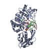
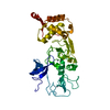
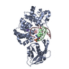
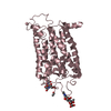
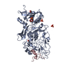
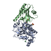
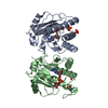
- PDB-4yph: Crystal Structure of MutY bound to its anti-substrate with the di... -
+ Open data

- Basic information
| Entry | Database: PDB / ID: 4yph | ||||||
|---|---|---|---|---|---|---|---|
| Title | Crystal Structure of MutY bound to its anti-substrate with the disulfide cross-linker reduced | ||||||
 Components Components |
| ||||||
 Keywords Keywords | Hydrolase/DNA / 8-oxoguanine / base-excision repair / anti-substrate / Hydrolase-DNA complex | ||||||
| Function / homology |  Function and homology information Function and homology informationadenine glycosylase / adenine/guanine mispair binding / DNA N-glycosylase activity / 8-oxo-7,8-dihydroguanine DNA N-glycosylase activity / purine-specific mismatch base pair DNA N-glycosylase activity / oxidized purine DNA binding / mismatch repair / base-excision repair / 4 iron, 4 sulfur cluster binding / DNA binding / metal ion binding Similarity search - Function | ||||||
| Biological species |   Geobacillus stearothermophilus (bacteria) Geobacillus stearothermophilus (bacteria)synthetic construct (others) | ||||||
| Method |  X-RAY DIFFRACTION / SYNCHROTRON / MOLECULAR REPLACEMENT / Resolution: 2.32 Å X-RAY DIFFRACTION / SYNCHROTRON / MOLECULAR REPLACEMENT / Resolution: 2.32 Å | ||||||
 Authors Authors | Wang, L. / Lee, S. / Verdine, G.L. | ||||||
 Citation Citation | Journal: J.Biol.Chem. / Year: 2015 Title: Structural Basis for Avoidance of Promutagenic DNA Repair by MutY Adenine DNA Glycosylase. Authors: Wang, L. / Lee, S.J. / Verdine, G.L. | ||||||
| History |
|
- Structure visualization
| Structure viewer | Molecule: MolmilJmol/JSmol |
|---|
- Downloads & links
-Download
| PDBx/mmCIF format | 4yph.cif.gz | 186.5 KB | Display | PDBx/mmCIF format |
|---|---|---|---|---|
| PDB format | pdb4yph.ent.gz | 142.3 KB | Display | PDB format |
| PDBx/mmJSON format | 4yph.json.gz | Tree view | PDBx/mmJSON format | |
| Others |  Other downloads Other downloads |
-Validation report
| Arichive directory | https://data.pdbj.org/pub/pdb/validation_reports/yp/4yphftp://data.pdbj.org/pub/pdb/validation_reports/yp/4yph | HTTPS FTP |
|---|
-Related structure data
| Related structure data |  4yoqC 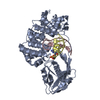 4yprC  1rrqS C: citing same article ( S: Starting model for refinement |
|---|---|
| Similar structure data |


-Links
 PDBj
PDBj


- Assembly
| Deposited unit | 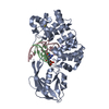
| ||||||||
|---|---|---|---|---|---|---|---|---|---|
| 1 |
| ||||||||
| Unit cell |
|
-Components
-Protein , 1 types, 1 molecules A
| #1: Protein | Mass: 42098.926 Da / Num. of mol.: 1 / Mutation: N144D, P164C Source method: isolated from a genetically manipulated source Source: (gene. exp.) Geobacillus stearothermophilus (bacteria)Plasmid: pET28 / Production host: References: UniProt: P83847, Hydrolases; Glycosylases; Hydrolysing N-glycosyl compounds |
|---|
-DNA chain , 2 types, 2 molecules BC
| #2: DNA chain | Mass: 3423.249 Da / Num. of mol.: 1 / Source method: obtained synthetically / Source: (synth.) synthetic construct (others) |
|---|---|
| #3: DNA chain | Mass: 3300.159 Da / Num. of mol.: 1 / Source method: obtained synthetically / Source: (synth.) synthetic construct (others) |
-Non-polymers , 3 types, 62 molecules 


| #4: Chemical | ChemComp-SF4 /  Mass: 351.640 Da / Num. of mol.: 1 / Source method: obtained synthetically / Formula: Fe4S4 Mass: 351.640 Da / Num. of mol.: 1 / Source method: obtained synthetically / Formula: Fe4S4 |
|---|---|
| #5: Chemical | ChemComp-NA /  Mass: 22.990 Da / Num. of mol.: 1 / Source method: obtained synthetically / Formula: Na Mass: 22.990 Da / Num. of mol.: 1 / Source method: obtained synthetically / Formula: Na |
| #6: Water | ChemComp-HOH / Mass: 18.015 Da / Num. of mol.: 60 / Source method: isolated from a natural source / Formula: H2O |
-Experimental details
-Experiment
| Experiment | Method: X-RAY DIFFRACTION / Number of used crystals: 1 |
|---|
- Sample preparation
| Crystal | Density Matthews: 2.3 Å3/Da / Density % sol: 46.61 % |
|---|---|
| Crystal grow | Temperature: 298 K / Method: vapor diffusion, hanging drop / pH: 8 Details: 100 mM Tris, pH 8.0, 250 mM NaOAc, 29% (wt/vol) PEG 4000, 25 mM TCEP |
-Data collection
| Diffraction | Mean temperature: 100 K |
|---|---|
| Diffraction source | Source: SYNCHROTRON / Site: APS  / Beamline: 24-ID-E / Wavelength: 0.98 Å / Beamline: 24-ID-E / Wavelength: 0.98 Å |
| Detector | Type: ADSC QUANTUM 315 / Detector: CCD / Date: Mar 13, 2014 |
| Radiation | Protocol: SINGLE WAVELENGTH / Monochromatic (M) / Laue (L): M / Scattering type: x-ray |
| Radiation wavelength | Wavelength: 0.98 Å / Relative weight: 1 |
| Reflection | Resolution: 2.32→83.76 Å / Num. obs: 20065 / % possible obs: 98.7 % / Redundancy: 3.5 % / Net I/σ(I): 8.8 |
| Reflection shell | Resolution: 2.32→2.45 Å / Redundancy: 3.7 % / Rmerge(I) obs: 0.855 / Mean I/σ(I) obs: 1.5 / % possible all: 100 |
- Processing
| Software |
| ||||||||||||||||||||||||||||||||||||||||||||||||||||||||
|---|---|---|---|---|---|---|---|---|---|---|---|---|---|---|---|---|---|---|---|---|---|---|---|---|---|---|---|---|---|---|---|---|---|---|---|---|---|---|---|---|---|---|---|---|---|---|---|---|---|---|---|---|---|---|---|---|---|
| Refinement | Method to determine structure: MOLECULAR REPLACEMENT Starting model: PDB entry 1RRQ Resolution: 2.32→41.37 Å / FOM work R set: 0.8162 / SU ML: 0.32 / Cross valid method: FREE R-VALUE / σ(F): 1.33 / Phase error: 24.81 / Stereochemistry target values: ML
| ||||||||||||||||||||||||||||||||||||||||||||||||||||||||
| Solvent computation | Shrinkage radii: 0.9 Å / VDW probe radii: 1.11 Å / Solvent model: FLAT BULK SOLVENT MODEL | ||||||||||||||||||||||||||||||||||||||||||||||||||||||||
| Displacement parameters | Biso max: 180.97 Å2 / Biso mean: 64.5 Å2 / Biso min: 20.93 Å2 | ||||||||||||||||||||||||||||||||||||||||||||||||||||||||
| Refinement step | Cycle: final / Resolution: 2.32→41.37 Å
| ||||||||||||||||||||||||||||||||||||||||||||||||||||||||
| Refine LS restraints |
| ||||||||||||||||||||||||||||||||||||||||||||||||||||||||
| LS refinement shell | Refine-ID: X-RAY DIFFRACTION / Total num. of bins used: 7
| ||||||||||||||||||||||||||||||||||||||||||||||||||||||||
| Refinement TLS params. | Method: refined / Origin x: -16.9774 Å / Origin y: 3.4422 Å / Origin z: -15.7758 Å
| ||||||||||||||||||||||||||||||||||||||||||||||||||||||||
| Refinement TLS group |
|
