 Movie
Movie Controller

[English] 日本語
 Yorodumi
- PDB-4xsh: The complex structure of C3cer exoenzyme and GTP bound RhoA (NADH... -
+ Open data

- Basic information
| Entry | Database: PDB / ID: 4xsh | ||||||
|---|---|---|---|---|---|---|---|
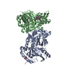
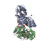
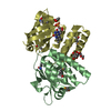
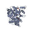
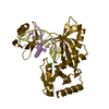
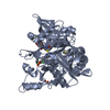
| Title | The complex structure of C3cer exoenzyme and GTP bound RhoA (NADH-bound state) | ||||||
 Components Components |
| ||||||
 Keywords Keywords | SIGNALING PROTEIN/Transferase / ADP Ribose Transferases / Bacterial Toxins / SIGNALING PROTEIN-Transferase complex | ||||||
| Function / homology |  Function and homology information Function and homology informationalpha-beta T cell lineage commitment / aortic valve formation / beta selection / positive regulation of lipase activity / endothelial tube lumen extension / skeletal muscle satellite cell migration / positive regulation of vascular associated smooth muscle contraction / angiotensin-mediated vasoconstriction involved in regulation of systemic arterial blood pressure / SLIT2:ROBO1 increases RHOA activity / RHO GTPases Activate Rhotekin and Rhophilins ...alpha-beta T cell lineage commitment / aortic valve formation / beta selection / positive regulation of lipase activity / endothelial tube lumen extension / skeletal muscle satellite cell migration / positive regulation of vascular associated smooth muscle contraction / angiotensin-mediated vasoconstriction involved in regulation of systemic arterial blood pressure / SLIT2:ROBO1 increases RHOA activity / RHO GTPases Activate Rhotekin and Rhophilins / Roundabout signaling pathway / bone trabecula morphogenesis / negative regulation of intracellular steroid hormone receptor signaling pathway / Axonal growth inhibition (RHOA activation) / Axonal growth stimulation / cleavage furrow formation / regulation of neural precursor cell proliferation / regulation of osteoblast proliferation / regulation of modification of postsynaptic actin cytoskeleton / forebrain radial glial cell differentiation / mitotic cleavage furrow formation / apical junction assembly / negative regulation of cell migration involved in sprouting angiogenesis / positive regulation of alpha-beta T cell differentiation / cell junction assembly / negative regulation of motor neuron apoptotic process / establishment of epithelial cell apical/basal polarity / cellular response to chemokine / regulation of systemic arterial blood pressure by endothelin / positive regulation of podosome assembly / negative regulation of oxidative phosphorylation / regulation of modification of postsynaptic structure / RHO GTPases Activate ROCKs / negative regulation of cell size / RHO GTPases activate CIT / odontogenesis / motor neuron apoptotic process / PCP/CE pathway / Sema4D induced cell migration and growth-cone collapse / RHO GTPases activate KTN1 / apolipoprotein A-I-mediated signaling pathway / wound healing, spreading of cells / Sema4D mediated inhibition of cell attachment and migration / positive regulation of leukocyte adhesion to vascular endothelial cell / Wnt signaling pathway, planar cell polarity pathway / PI3K/AKT activation / ossification involved in bone maturation / regulation of focal adhesion assembly / androgen receptor signaling pathway / negative chemotaxis / EPHA-mediated growth cone collapse / apical junction complex / stress fiber assembly / myosin binding / regulation of neuron projection development / positive regulation of cytokinesis / RHOC GTPase cycle / cellular response to cytokine stimulus / cerebral cortex cell migration / ERBB2 Regulates Cell Motility / positive regulation of protein serine/threonine kinase activity / cleavage furrow / semaphorin-plexin signaling pathway / negative regulation of cell-substrate adhesion / ficolin-1-rich granule membrane / mitotic spindle assembly / RHOA GTPase cycle / positive regulation of T cell migration / endothelial cell migration / skeletal muscle tissue development / Rho protein signal transduction / RHO GTPases activate PKNs / GPVI-mediated activation cascade / PTK6 Regulates RHO GTPases, RAS GTPase and MAP kinases / negative regulation of reactive oxygen species biosynthetic process / substrate adhesion-dependent cell spreading / positive regulation of stress fiber assembly / cytoplasmic microtubule organization / EPHB-mediated forward signaling / positive regulation of neuron differentiation / substantia nigra development / regulation of cell migration / secretory granule membrane / cell-matrix adhesion / kidney development / cell periphery / regulation of microtubule cytoskeleton organization / regulation of actin cytoskeleton organization / small monomeric GTPase / TGF-beta receptor signaling in EMT (epithelial to mesenchymal transition) / RHO GTPases Activate Formins / positive regulation of non-canonical NF-kappaB signal transduction / VEGFA-VEGFR2 Pathway / neuron migration / cell morphogenesis / ruffle membrane / cytoplasmic side of plasma membrane / cell junction / Ovarian tumor domain proteases / G beta:gamma signalling through PI3Kgamma Similarity search - Function | ||||||
| Biological species |  Homo sapiens (human) Homo sapiens (human) | ||||||
| Method |  X-RAY DIFFRACTION / MOLECULAR REPLACEMENT / Resolution: 2.5 Å X-RAY DIFFRACTION / MOLECULAR REPLACEMENT / Resolution: 2.5 Å | ||||||
 Authors Authors | Toda, A. / Tsurumura, T. / Yoshida, T. / Tsuge, H. | ||||||
| Funding support |  Japan, 1items Japan, 1items
| ||||||
 Citation Citation | Journal: J.Biol.Chem. / Year: 2015 Title: Rho GTPase Recognition by C3 Exoenzyme Based on C3-RhoA Complex Structure. Authors: Toda, A. / Tsurumura, T. / Yoshida, T. / Tsumori, Y. / Tsuge, H. | ||||||
| History |
|
- Structure visualization
| Structure viewer | Molecule: MolmilJmol/JSmol |
|---|
- Downloads & links
-Download
| PDBx/mmCIF format | 4xsh.cif.gz | 174.7 KB | Display | PDBx/mmCIF format |
|---|---|---|---|---|
| PDB format | pdb4xsh.ent.gz | 137.4 KB | Display | PDB format |
| PDBx/mmJSON format | 4xsh.json.gz | Tree view | PDBx/mmJSON format | |
| Others |  Other downloads Other downloads |
-Validation report
| Arichive directory | https://data.pdbj.org/pub/pdb/validation_reports/xs/4xshftp://data.pdbj.org/pub/pdb/validation_reports/xs/4xsh | HTTPS FTP |
|---|
-Related structure data
| Related structure data |  4xsgC  5bwmC 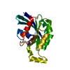 1a2bS 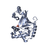 3bw8S C: citing same article ( S: Starting model for refinement |
|---|---|
| Similar structure data |


-Links
 PDBj
PDBj


- Assembly
| Deposited unit | 
| ||||||||
|---|---|---|---|---|---|---|---|---|---|
| 1 |
| ||||||||
| Unit cell |
|
-Components
-Protein , 2 types, 2 molecules AB
| #1: Protein | Mass: 20235.180 Da / Num. of mol.: 1 / Fragment: UNP residues 1-179 / Mutation: F25N Source method: isolated from a genetically manipulated source Source: (gene. exp.) Homo sapiens (human) / Gene: RHOA, ARH12, ARHA, RHO12 / Plasmid: pGEX4T-1 / Production host: |
|---|---|
| #2: Protein | Mass: 25288.775 Da / Num. of mol.: 1 Source method: isolated from a genetically manipulated source Source: (gene. exp.) |
-Non-polymers , 5 types, 61 molecules 


| #3: Chemical | ChemComp-GSP /  Mass: 539.246 Da / Num. of mol.: 1 / Source method: obtained synthetically / Formula: C10H16N5O13P3S Mass: 539.246 Da / Num. of mol.: 1 / Source method: obtained synthetically / Formula: C10H16N5O13P3S | ||
|---|---|---|---|
| #4: Chemical | ChemComp-MG /  Mass: 24.305 Da / Num. of mol.: 1 / Source method: obtained synthetically / Formula: Mg Mass: 24.305 Da / Num. of mol.: 1 / Source method: obtained synthetically / Formula: Mg | ||
| #5: Chemical | ChemComp-NAI /  Mass: 665.441 Da / Num. of mol.: 1 / Source method: obtained synthetically / Formula: C21H29N7O14P2 Mass: 665.441 Da / Num. of mol.: 1 / Source method: obtained synthetically / Formula: C21H29N7O14P2 | ||
| #6: Chemical |  Mass: 62.068 Da / Num. of mol.: 3 / Source method: obtained synthetically / Formula: C2H6O2 Mass: 62.068 Da / Num. of mol.: 3 / Source method: obtained synthetically / Formula: C2H6O2#7: Water | ChemComp-HOH / | Mass: 18.015 Da / Num. of mol.: 55 / Source method: isolated from a natural source / Formula: H2O |
-Experimental details
-Experiment
| Experiment | Method: X-RAY DIFFRACTION / Number of used crystals: 1 |
|---|
- Sample preparation
| Crystal | Density Matthews: 2.28 Å3/Da / Density % sol: 46.08 % |
|---|---|
| Crystal grow | Temperature: 277 K / Method: vapor diffusion, hanging drop / Details: 100 mM MES (pH 6.4), 20% PEG1500 |
-Data collection
| Diffraction | Mean temperature: 100 K |
|---|---|
| Diffraction source | Source: ROTATING ANODE / Type: RIGAKU MICROMAX-007 HF / Wavelength: 1.54 Å |
| Detector | Type: RIGAKU RAXIS VII / Detector: IMAGE PLATE / Date: Sep 30, 2014 |
| Radiation | Protocol: SINGLE WAVELENGTH / Monochromatic (M) / Laue (L): M / Scattering type: x-ray |
| Radiation wavelength | Wavelength: 1.54 Å / Relative weight: 1 |
| Reflection | Resolution: 2.3→50 Å / Num. obs: 13464 / % possible obs: 99.9 % / Redundancy: 5.5 % / Net I/σ(I): 21 |
- Processing
| Software |
| ||||||||||||||||||||||||||||||||||||||||||||||||||||||||||||||||||||||||||||||||||||||||||||||||||||||||||||||||||||||||||||||||||||||||||||||||||||||||||||||||||||||||||||||||||||||
|---|---|---|---|---|---|---|---|---|---|---|---|---|---|---|---|---|---|---|---|---|---|---|---|---|---|---|---|---|---|---|---|---|---|---|---|---|---|---|---|---|---|---|---|---|---|---|---|---|---|---|---|---|---|---|---|---|---|---|---|---|---|---|---|---|---|---|---|---|---|---|---|---|---|---|---|---|---|---|---|---|---|---|---|---|---|---|---|---|---|---|---|---|---|---|---|---|---|---|---|---|---|---|---|---|---|---|---|---|---|---|---|---|---|---|---|---|---|---|---|---|---|---|---|---|---|---|---|---|---|---|---|---|---|---|---|---|---|---|---|---|---|---|---|---|---|---|---|---|---|---|---|---|---|---|---|---|---|---|---|---|---|---|---|---|---|---|---|---|---|---|---|---|---|---|---|---|---|---|---|---|---|---|---|
| Refinement | Method to determine structure: MOLECULAR REPLACEMENT Starting model: 1A2B and 3BW8 Resolution: 2.5→45.56 Å / Cor.coef. Fo:Fc: 0.963 / Cor.coef. Fo:Fc free: 0.93 / SU B: 27.386 / SU ML: 0.294 / Cross valid method: THROUGHOUT / ESU R Free: 0.339 / Stereochemistry target values: MAXIMUM LIKELIHOOD / Details: HYDROGENS HAVE BEEN ADDED IN THE RIDING POSITIONS
| ||||||||||||||||||||||||||||||||||||||||||||||||||||||||||||||||||||||||||||||||||||||||||||||||||||||||||||||||||||||||||||||||||||||||||||||||||||||||||||||||||||||||||||||||||||||
| Solvent computation | Ion probe radii: 0.8 Å / Shrinkage radii: 0.8 Å / VDW probe radii: 1.2 Å / Solvent model: MASK | ||||||||||||||||||||||||||||||||||||||||||||||||||||||||||||||||||||||||||||||||||||||||||||||||||||||||||||||||||||||||||||||||||||||||||||||||||||||||||||||||||||||||||||||||||||||
| Displacement parameters | Biso mean: 68.204 Å2
| ||||||||||||||||||||||||||||||||||||||||||||||||||||||||||||||||||||||||||||||||||||||||||||||||||||||||||||||||||||||||||||||||||||||||||||||||||||||||||||||||||||||||||||||||||||||
| Refinement step | Cycle: LAST / Resolution: 2.5→45.56 Å
| ||||||||||||||||||||||||||||||||||||||||||||||||||||||||||||||||||||||||||||||||||||||||||||||||||||||||||||||||||||||||||||||||||||||||||||||||||||||||||||||||||||||||||||||||||||||
| Refine LS restraints |
|