 Movie
Movie Controller


+ Open data

- Basic information
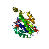
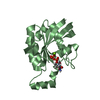
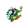
| Entry | Database: PDB / ID: 1a2b | ||||||
|---|---|---|---|---|---|---|---|
| Title | HUMAN RHOA COMPLEXED WITH GTP ANALOGUE | ||||||
 Components Components | TRANSFORMING PROTEIN RHOA | ||||||
 Keywords Keywords | ONCOGENE PROTEIN / SMALL G-PROTEIN / SIGNAL TRANSDUCTION / GTPASE / RAS SUPERFAMILY | ||||||
| Function / homology |  Function and homology information Function and homology informationalpha-beta T cell lineage commitment / aortic valve formation / beta selection / positive regulation of lipase activity / endothelial tube lumen extension / skeletal muscle satellite cell migration / positive regulation of vascular associated smooth muscle contraction / angiotensin-mediated vasoconstriction involved in regulation of systemic arterial blood pressure / SLIT2:ROBO1 increases RHOA activity / RHO GTPases Activate Rhotekin and Rhophilins ...alpha-beta T cell lineage commitment / aortic valve formation / beta selection / positive regulation of lipase activity / endothelial tube lumen extension / skeletal muscle satellite cell migration / positive regulation of vascular associated smooth muscle contraction / angiotensin-mediated vasoconstriction involved in regulation of systemic arterial blood pressure / SLIT2:ROBO1 increases RHOA activity / RHO GTPases Activate Rhotekin and Rhophilins / Roundabout signaling pathway / bone trabecula morphogenesis / negative regulation of intracellular steroid hormone receptor signaling pathway / Axonal growth inhibition (RHOA activation) / Axonal growth stimulation / cleavage furrow formation / regulation of neural precursor cell proliferation / regulation of osteoblast proliferation / regulation of modification of postsynaptic actin cytoskeleton / forebrain radial glial cell differentiation / mitotic cleavage furrow formation / apical junction assembly / negative regulation of cell migration involved in sprouting angiogenesis / positive regulation of alpha-beta T cell differentiation / cell junction assembly / negative regulation of motor neuron apoptotic process / establishment of epithelial cell apical/basal polarity / cellular response to chemokine / regulation of systemic arterial blood pressure by endothelin / positive regulation of podosome assembly / negative regulation of oxidative phosphorylation / regulation of modification of postsynaptic structure / RHO GTPases Activate ROCKs / negative regulation of cell size / RHO GTPases activate CIT / odontogenesis / motor neuron apoptotic process / PCP/CE pathway / Sema4D induced cell migration and growth-cone collapse / RHO GTPases activate KTN1 / apolipoprotein A-I-mediated signaling pathway / wound healing, spreading of cells / Sema4D mediated inhibition of cell attachment and migration / positive regulation of leukocyte adhesion to vascular endothelial cell / Wnt signaling pathway, planar cell polarity pathway / PI3K/AKT activation / ossification involved in bone maturation / regulation of focal adhesion assembly / androgen receptor signaling pathway / negative chemotaxis / EPHA-mediated growth cone collapse / apical junction complex / stress fiber assembly / myosin binding / regulation of neuron projection development / positive regulation of cytokinesis / RHOC GTPase cycle / cellular response to cytokine stimulus / cerebral cortex cell migration / ERBB2 Regulates Cell Motility / positive regulation of protein serine/threonine kinase activity / cleavage furrow / semaphorin-plexin signaling pathway / negative regulation of cell-substrate adhesion / ficolin-1-rich granule membrane / mitotic spindle assembly / RHOA GTPase cycle / positive regulation of T cell migration / endothelial cell migration / skeletal muscle tissue development / Rho protein signal transduction / RHO GTPases activate PKNs / GPVI-mediated activation cascade / PTK6 Regulates RHO GTPases, RAS GTPase and MAP kinases / negative regulation of reactive oxygen species biosynthetic process / substrate adhesion-dependent cell spreading / positive regulation of stress fiber assembly / cytoplasmic microtubule organization / EPHB-mediated forward signaling / positive regulation of neuron differentiation / substantia nigra development / regulation of cell migration / secretory granule membrane / cell-matrix adhesion / kidney development / cell periphery / regulation of microtubule cytoskeleton organization / regulation of actin cytoskeleton organization / small monomeric GTPase / TGF-beta receptor signaling in EMT (epithelial to mesenchymal transition) / RHO GTPases Activate Formins / positive regulation of non-canonical NF-kappaB signal transduction / VEGFA-VEGFR2 Pathway / neuron migration / cell morphogenesis / ruffle membrane / cytoplasmic side of plasma membrane / cell junction / Ovarian tumor domain proteases / G beta:gamma signalling through PI3Kgamma Similarity search - Function | ||||||
| Biological species |  Homo sapiens (human) Homo sapiens (human) | ||||||
| Method |  X-RAY DIFFRACTION / MOLECULAR REPLACEMENT / Resolution: 2.4 Å X-RAY DIFFRACTION / MOLECULAR REPLACEMENT / Resolution: 2.4 Å | ||||||
 Authors Authors | Ihara, K. / Muraguchi, S. / Kato, M. / Shimizu, T. / Shirakawa, M. / Kuroda, S. / Kaibuchi, K. / Hakoshima, T. | ||||||
 Citation Citation | Journal: J.Biol.Chem. / Year: 1998 Title: Crystal structure of human RhoA in a dominantly active form complexed with a GTP analogue. Authors: Ihara, K. / Muraguchi, S. / Kato, M. / Shimizu, T. / Shirakawa, M. / Kuroda, S. / Kaibuchi, K. / Hakoshima, T. #1: Journal: Embo J. / Year: 1990Title: Refined Crystal Structure of the Triphosphate Conformation of H-Ras P21 at 1.35 A Resolution: Implications for the Mechanism of GTP Hydrolysis Authors: Pai, E.F. / Krengel, U. / Petsko, G.A. / Goody, R.S. / Kabsch, W. / Wittinghofer, A. | ||||||
| History |
|
- Structure visualization
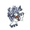
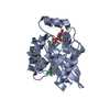
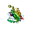
| Structure viewer | Molecule: MolmilJmol/JSmol |
|---|
- Downloads & links
-Download
| PDBx/mmCIF format | 1a2b.cif.gz | 50.8 KB | Display | PDBx/mmCIF format |
|---|---|---|---|---|
| PDB format | pdb1a2b.ent.gz | 35.4 KB | Display | PDB format |
| PDBx/mmJSON format | 1a2b.json.gz | Tree view | PDBx/mmJSON format | |
| Others |  Other downloads Other downloads |
-Validation report
| Arichive directory | https://data.pdbj.org/pub/pdb/validation_reports/a2/1a2bftp://data.pdbj.org/pub/pdb/validation_reports/a2/1a2b | HTTPS FTP |
|---|
-Related structure data
| Related structure data | 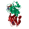 5p21S S: Starting model for refinement |
|---|---|
| Similar structure data |


-Links
 PDBj
PDBj


- Assembly
| Deposited unit | 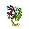
| ||||||||
|---|---|---|---|---|---|---|---|---|---|
| 1 |
| ||||||||
| Unit cell |
|
-Components
| #1: Protein | Mass: 20596.615 Da / Num. of mol.: 1 / Fragment: RESIDUES 1 - 181 Mutation: G14V, RESIDUES 1 - 181 WERE CLONED, THE N-TERMINUS CONTAINS A HIS-TAG Source method: isolated from a genetically manipulated source Details: COMPLEXED WITH ONE GTPGAMMAS AND ONE MG ION / Source: (gene. exp.) Homo sapiens (human) / Plasmid: PRSET B (INVITROGEN CO.) / Species (production host): Escherichia coli / Cellular location (production host): CYTOSOL / Production host:  |
|---|---|
| #2: Chemical | ChemComp-MG /   Mass: 24.305 Da / Num. of mol.: 1 / Source method: obtained synthetically / Formula: Mg Mass: 24.305 Da / Num. of mol.: 1 / Source method: obtained synthetically / Formula: Mg |
| #3: Chemical | ChemComp-GSP /   Mass: 539.246 Da / Num. of mol.: 1 / Source method: obtained synthetically / Formula: C10H16N5O13P3S Mass: 539.246 Da / Num. of mol.: 1 / Source method: obtained synthetically / Formula: C10H16N5O13P3S |
| #4: Water | ChemComp-HOH /  Mass: 18.015 Da / Num. of mol.: 38 / Source method: isolated from a natural source / Formula: H2O Mass: 18.015 Da / Num. of mol.: 38 / Source method: isolated from a natural source / Formula: H2O |
-Experimental details
-Experiment
| Experiment | Method: X-RAY DIFFRACTION / Number of used crystals: 1 |
|---|
- Sample preparation
| Crystal | Density Matthews: 2.77 Å3/Da / Density % sol: 56 % | ||||||||||||||||||||||||||||||||||||||||
|---|---|---|---|---|---|---|---|---|---|---|---|---|---|---|---|---|---|---|---|---|---|---|---|---|---|---|---|---|---|---|---|---|---|---|---|---|---|---|---|---|---|
| Crystal grow | Temperature: 277 K / Method: vapor diffusion, hanging drop / pH: 8.5 Details: CRYSTALS WERE OBTAINED AT 277 K BY THE HANGING-DROP VAPOR DIFFUSION METHOD FROM SOLUTIONS CONTAINING 10 MG/ML(PROTEIN,GTPGAMMAS,MG2+ MIXTURE), 10% PEG 8000,7.5% 14-DIOXANE, 50 MM TRIS-HCL PH ...Details: CRYSTALS WERE OBTAINED AT 277 K BY THE HANGING-DROP VAPOR DIFFUSION METHOD FROM SOLUTIONS CONTAINING 10 MG/ML(PROTEIN,GTPGAMMAS,MG2+ MIXTURE), 10% PEG 8000,7.5% 14-DIOXANE, 50 MM TRIS-HCL PH 8.5, EQUILIBRATED AGAINST 20% PEG 8000,15% 14-DIOXANE, 100 MM TRIS-HCL PH 8.5, vapor diffusion - hanging drop PH range: 7.5-8.5 | ||||||||||||||||||||||||||||||||||||||||
| Crystal grow | *PLUS Temperature: 4 ℃ / Method: vapor diffusion, hanging drop | ||||||||||||||||||||||||||||||||||||||||
| Components of the solutions | *PLUS
|
-Data collection
| Diffraction | Mean temperature: 283 K |
|---|---|
| Diffraction source | Source: ROTATING ANODE / Type: RIGAKU RUH3R / Wavelength: 1.5418 |
| Detector | Type: RIGAKU RAXIS IIC / Detector: IMAGE PLATE / Date: Mar 1, 1997 |
| Radiation | Monochromator: GRAPHITE(002) / Monochromatic (M) / Laue (L): M / Scattering type: x-ray |
| Radiation wavelength | Wavelength: 1.5418 Å / Relative weight: 1 |
| Reflection | Highest resolution: 2.4 Å / Num. obs: 8683 / % possible obs: 89.3 % / Observed criterion σ(I): 1 / Biso Wilson estimate: 31.6 Å2 / Rmerge(I) obs: 0.0875 / Net I/σ(I): 7.91 |
| Reflection shell | Resolution: 2.4→2.5 Å / Rmerge(I) obs: 0.267 / Mean I/σ(I) obs: 2.04 / % possible all: 74.8 |
| Reflection | *PLUS Num. measured all: 61579 |
| Reflection shell | *PLUS % possible obs: 74.8 % |
- Processing
| Software |
| ||||||||||||||||||||||||||||||||||||||||||||||||||||||||||||
|---|---|---|---|---|---|---|---|---|---|---|---|---|---|---|---|---|---|---|---|---|---|---|---|---|---|---|---|---|---|---|---|---|---|---|---|---|---|---|---|---|---|---|---|---|---|---|---|---|---|---|---|---|---|---|---|---|---|---|---|---|---|
| Refinement | Method to determine structure: MOLECULAR REPLACEMENT Starting model: PDB ENTRY 5P21 Resolution: 2.4→15 Å / Data cutoff high absF: 10000000 / Data cutoff low absF: 0.0001 / Cross valid method: THROUGHOUT / σ(F): 1
| ||||||||||||||||||||||||||||||||||||||||||||||||||||||||||||
| Displacement parameters | Biso mean: 43.5 Å2 | ||||||||||||||||||||||||||||||||||||||||||||||||||||||||||||
| Refinement step | Cycle: LAST / Resolution: 2.4→15 Å
| ||||||||||||||||||||||||||||||||||||||||||||||||||||||||||||
| Refine LS restraints |
| ||||||||||||||||||||||||||||||||||||||||||||||||||||||||||||
| LS refinement shell | Resolution: 2.4→2.51 Å / Total num. of bins used: 8
| ||||||||||||||||||||||||||||||||||||||||||||||||||||||||||||
| Xplor file |
| ||||||||||||||||||||||||||||||||||||||||||||||||||||||||||||
| Software | *PLUS Name: X-PLOR / Version: 3.8 / Classification: refinement | ||||||||||||||||||||||||||||||||||||||||||||||||||||||||||||
| Refinement | *PLUS | ||||||||||||||||||||||||||||||||||||||||||||||||||||||||||||
| Solvent computation | *PLUS | ||||||||||||||||||||||||||||||||||||||||||||||||||||||||||||
| Displacement parameters | *PLUS | ||||||||||||||||||||||||||||||||||||||||||||||||||||||||||||
| Refine LS restraints | *PLUS
| ||||||||||||||||||||||||||||||||||||||||||||||||||||||||||||
| LS refinement shell | *PLUS Rfactor obs: 0.306 |
