 Movie
Movie Controller


+ Open data

- Basic information
| Entry | Database: PDB / ID: 4h03 | ||||||
|---|---|---|---|---|---|---|---|
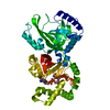
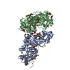
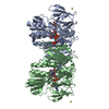
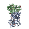
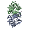
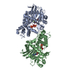
| Title | Crystal structure of NAD+-Ia-actin complex | ||||||
 Components Components |
| ||||||
 Keywords Keywords | TOXIN/STRUCTURAL PROTEIN / ADP-ribosyltransferase / TOXIN-STRUCTURAL PROTEIN complex | ||||||
| Function / homology |  Function and homology information Function and homology informationcytoskeletal motor activator activity / myosin heavy chain binding / tropomyosin binding / actin filament bundle / troponin I binding / filamentous actin / mesenchyme migration / skeletal muscle myofibril / actin filament bundle assembly / striated muscle thin filament ...cytoskeletal motor activator activity / myosin heavy chain binding / tropomyosin binding / actin filament bundle / troponin I binding / filamentous actin / mesenchyme migration / skeletal muscle myofibril / actin filament bundle assembly / striated muscle thin filament / skeletal muscle thin filament assembly / actin monomer binding / skeletal muscle fiber development / stress fiber / titin binding / actin filament polymerization / filopodium / actin filament / Hydrolases; Acting on acid anhydrides; Acting on acid anhydrides to facilitate cellular and subcellular movement / calcium-dependent protein binding / lamellipodium / cell body / protein domain specific binding / nucleotide binding / hydrolase activity / calcium ion binding / positive regulation of gene expression / magnesium ion binding / extracellular region / ATP binding / identical protein binding / cytoplasm Similarity search - Function | ||||||
| Biological species |   Clostridium perfringens (bacteria) Clostridium perfringens (bacteria) | ||||||
| Method |  X-RAY DIFFRACTION / SYNCHROTRON / MOLECULAR REPLACEMENT / Resolution: 1.75 Å X-RAY DIFFRACTION / SYNCHROTRON / MOLECULAR REPLACEMENT / Resolution: 1.75 Å | ||||||
 Authors Authors | Tsurumura, T. / Oda, M. / Nagahama, M. / Tsuge, H. | ||||||
 Citation Citation | Journal: Proc.Natl.Acad.Sci.USA / Year: 2013 Title: Arginine ADP-ribosylation mechanism based on structural snapshots of iota-toxin and actin complex Authors: Tsurumura, T. / Tsumori, Y. / Qiu, H. / Oda, M. / Sakurai, J. / Nagahama, M. / Tsuge, H. | ||||||
| History |
|
- Structure visualization
| Structure viewer | Molecule: MolmilJmol/JSmol |
|---|
- Downloads & links
-Download
| PDBx/mmCIF format | 4h03.cif.gz | 322.4 KB | Display | PDBx/mmCIF format |
|---|---|---|---|---|
| PDB format | pdb4h03.ent.gz | 257.5 KB | Display | PDB format |
| PDBx/mmJSON format | 4h03.json.gz | Tree view | PDBx/mmJSON format | |
| Others |  Other downloads Other downloads |
-Validation report
| Arichive directory | https://data.pdbj.org/pub/pdb/validation_reports/h0/4h03ftp://data.pdbj.org/pub/pdb/validation_reports/h0/4h03 | HTTPS FTP |
|---|
-Related structure data
| Related structure data | 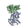 4gy2C 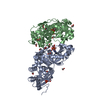 4h0tC  4h0vC 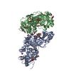 4h0xC  4h0yC 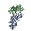 3buzS S: Starting model for refinement C: citing same article ( |
|---|---|
| Similar structure data |


-Links
 PDBj
PDBj


- Assembly
| Deposited unit | 
| ||||||||
|---|---|---|---|---|---|---|---|---|---|
| 1 |
| ||||||||
| Unit cell |
|
-Components
-Protein , 2 types, 2 molecules AB
| #1: Protein | Mass: 48219.180 Da / Num. of mol.: 1 Source method: isolated from a genetically manipulated source Source: (gene. exp.) Clostridium perfringens (bacteria) / Production host: References: UniProt: Q46220, NAD+-protein-arginine ADP-ribosyltransferase |
|---|---|
| #2: Protein | Mass: 41875.633 Da / Num. of mol.: 1 Source method: isolated from a genetically manipulated source Source: (gene. exp.) |
-Non-polymers , 7 types, 325 molecules 


| #3: Chemical | ChemComp-NAD /  Mass: 663.425 Da / Num. of mol.: 1 / Source method: obtained synthetically / Formula: C21H27N7O14P2 / Comment: NAD*YM Mass: 663.425 Da / Num. of mol.: 1 / Source method: obtained synthetically / Formula: C21H27N7O14P2 / Comment: NAD*YM | ||||||||
|---|---|---|---|---|---|---|---|---|---|
| #4: Chemical | ChemComp-PO4 /  Mass: 94.971 Da / Num. of mol.: 1 / Source method: obtained synthetically / Formula: PO4 Mass: 94.971 Da / Num. of mol.: 1 / Source method: obtained synthetically / Formula: PO4 | ||||||||
| #5: Chemical | ChemComp-EDO /  Mass: 62.068 Da / Num. of mol.: 48 / Source method: obtained synthetically / Formula: C2H6O2 Mass: 62.068 Da / Num. of mol.: 48 / Source method: obtained synthetically / Formula: C2H6O2#6: Chemical | ChemComp-CA / |  Mass: 40.078 Da / Num. of mol.: 1 / Source method: obtained synthetically / Formula: Ca Mass: 40.078 Da / Num. of mol.: 1 / Source method: obtained synthetically / Formula: Ca#7: Chemical | ChemComp-ATP / |  Mass: 507.181 Da / Num. of mol.: 1 / Source method: obtained synthetically / Formula: C10H16N5O13P3 / Comment: ATP, energy-carrying molecule*YM Mass: 507.181 Da / Num. of mol.: 1 / Source method: obtained synthetically / Formula: C10H16N5O13P3 / Comment: ATP, energy-carrying molecule*YM#8: Chemical | ChemComp-LAR / |  Mass: 421.550 Da / Num. of mol.: 1 / Source method: obtained synthetically / Formula: C22H31NO5S / Comment: toxin*YM Mass: 421.550 Da / Num. of mol.: 1 / Source method: obtained synthetically / Formula: C22H31NO5S / Comment: toxin*YM#9: Water | ChemComp-HOH / | Mass: 18.015 Da / Num. of mol.: 272 / Source method: isolated from a natural source / Formula: H2O |
-Experimental details
-Experiment
| Experiment | Method: X-RAY DIFFRACTION / Number of used crystals: 1 |
|---|
- Sample preparation
| Crystal | Density Matthews: 3.12 Å3/Da / Density % sol: 60.62 % |
|---|---|
| Crystal grow | Temperature: 277.13 K / Method: vapor diffusion, hanging drop / pH: 6.5 Details: 18% PEG1500, 0.1M MES, pH 6.5, VAPOR DIFFUSION, HANGING DROP, temperature 277.13K |
-Data collection
| Diffraction | Mean temperature: 100 K |
|---|---|
| Diffraction source | Source: SYNCHROTRON / Site: Photon Factory  / Beamline: BL-5A / Wavelength: 1 Å / Beamline: BL-5A / Wavelength: 1 Å |
| Detector | Type: ADSC QUANTUM 210r / Detector: CCD / Date: May 29, 2012 |
| Radiation | Protocol: SINGLE WAVELENGTH / Monochromatic (M) / Laue (L): M / Scattering type: x-ray |
| Radiation wavelength | Wavelength: 1 Å / Relative weight: 1 |
| Reflection | Resolution: 1.75→50 Å / Num. all: 114683 / Num. obs: 110554 / % possible obs: 96.4 % / Observed criterion σ(F): 0 / Observed criterion σ(I): 0 / Redundancy: 5.5 % / Rmerge(I) obs: 0.048 / Net I/σ(I): 19.9 |
| Reflection shell | Resolution: 1.75→1.78 Å / Redundancy: 3.6 % / Rmerge(I) obs: 0.416 / Mean I/σ(I) obs: 3.2 / Num. unique all: 5154 / % possible all: 90.9 |
- Processing
| Software |
| |||||||||||||||||||||||||||||||||||||||||||||||||||||||||||||||||||||||||||
|---|---|---|---|---|---|---|---|---|---|---|---|---|---|---|---|---|---|---|---|---|---|---|---|---|---|---|---|---|---|---|---|---|---|---|---|---|---|---|---|---|---|---|---|---|---|---|---|---|---|---|---|---|---|---|---|---|---|---|---|---|---|---|---|---|---|---|---|---|---|---|---|---|---|---|---|---|
| Refinement | Method to determine structure: MOLECULAR REPLACEMENT Starting model: PDB ENTRY 3BUZ Resolution: 1.75→26.29 Å / Cor.coef. Fo:Fc: 0.946 / Cor.coef. Fo:Fc free: 0.938 / SU B: 4.187 / SU ML: 0.066 / Cross valid method: THROUGHOUT / ESU R: 0.115 / ESU R Free: 0.107 / Stereochemistry target values: MAXIMUM LIKELIHOOD / Details: HYDROGENS HAVE BEEN ADDED IN THE RIDING POSITIONS
| |||||||||||||||||||||||||||||||||||||||||||||||||||||||||||||||||||||||||||
| Solvent computation | Ion probe radii: 0.8 Å / Shrinkage radii: 0.8 Å / VDW probe radii: 1.2 Å / Solvent model: MASK | |||||||||||||||||||||||||||||||||||||||||||||||||||||||||||||||||||||||||||
| Displacement parameters | Biso mean: 29.68 Å2
| |||||||||||||||||||||||||||||||||||||||||||||||||||||||||||||||||||||||||||
| Refinement step | Cycle: LAST / Resolution: 1.75→26.29 Å
| |||||||||||||||||||||||||||||||||||||||||||||||||||||||||||||||||||||||||||
| Refine LS restraints |
| |||||||||||||||||||||||||||||||||||||||||||||||||||||||||||||||||||||||||||
| LS refinement shell | Resolution: 1.748→1.794 Å / Total num. of bins used: 20
| |||||||||||||||||||||||||||||||||||||||||||||||||||||||||||||||||||||||||||
| Refinement TLS params. | Method: refined / Refine-ID: X-RAY DIFFRACTION
| |||||||||||||||||||||||||||||||||||||||||||||||||||||||||||||||||||||||||||
| Refinement TLS group |
|
