 Movie
Movie Controller

[English] 日本語
 Yorodumi
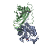
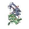
- PDB-1giq: Crystal Structure of the Enzymatic Componet of Iota-Toxin from Cl... -
+ Open data

- Basic information
| Entry | Database: PDB / ID: 1giq | ||||||
|---|---|---|---|---|---|---|---|
| Title | Crystal Structure of the Enzymatic Componet of Iota-Toxin from Clostridium Perfringens with NADH | ||||||
 Components Components | IOTA TOXIN COMPONENT IA | ||||||
 Keywords Keywords | TOXIN / enzymatic component | ||||||
| Function / homology |  Function and homology information Function and homology information | ||||||
| Biological species |   Clostridium perfringens (bacteria) Clostridium perfringens (bacteria) | ||||||
| Method |  X-RAY DIFFRACTION / SYNCHROTRON / MOLECULAR REPLACEMENT / Resolution: 1.8 Å X-RAY DIFFRACTION / SYNCHROTRON / MOLECULAR REPLACEMENT / Resolution: 1.8 Å | ||||||
 Authors Authors | Tsuge, H. / Nagahama, M. / Nishimura, H. / Hisatsune, J. / Sakaguchi, Y. / Itogawa, Y. / Katunuma, N. / Sakurai, J. | ||||||
 Citation Citation | Journal: J.MOL.BIOL. / Year: 2003 Title: Crystal Structure and Site-directed Mutagenesis of Enzymatic Components from Clostridium perfringens Iota-toxin Authors: Tsuge, H. / Nagahama, M. / Nishimura, H. / Hisatsune, J. / Sakaguchi, Y. / Itogawa, Y. / Katunuma, N. / Sakurai, J. | ||||||
| History |
|
- Structure visualization
| Structure viewer | Molecule: MolmilJmol/JSmol |
|---|
- Downloads & links
-Download
| PDBx/mmCIF format | 1giq.cif.gz | 184.2 KB | Display | PDBx/mmCIF format |
|---|---|---|---|---|
| PDB format | pdb1giq.ent.gz | 145.3 KB | Display | PDB format |
| PDBx/mmJSON format | 1giq.json.gz | Tree view | PDBx/mmJSON format | |
| Others |  Other downloads Other downloads |
-Validation report
| Arichive directory | https://data.pdbj.org/pub/pdb/validation_reports/gi/1giqftp://data.pdbj.org/pub/pdb/validation_reports/gi/1giq | HTTPS FTP |
|---|
-Related structure data
| Related structure data |  1girSC S: Starting model for refinement C: citing same article ( |
|---|---|
| Similar structure data |


-Links
 PDBj
PDBj

- Assembly
| Deposited unit | 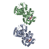
| ||||||||
|---|---|---|---|---|---|---|---|---|---|
| 1 | 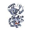
| ||||||||
| 2 | 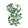
| ||||||||
| Unit cell |
|
-Components
| #1: Protein | Mass: 47648.516 Da / Num. of mol.: 2 / Source method: isolated from a natural source / Source: (natural) Clostridium perfringens (bacteria) / References: UniProt: Q46220#2: Chemical |   Mass: 665.441 Da / Num. of mol.: 2 / Source method: obtained synthetically / Formula: C21H29N7O14P2 Mass: 665.441 Da / Num. of mol.: 2 / Source method: obtained synthetically / Formula: C21H29N7O14P2#3: Water | ChemComp-HOH / |  Mass: 18.015 Da / Num. of mol.: 286 / Source method: isolated from a natural source / Formula: H2O Mass: 18.015 Da / Num. of mol.: 286 / Source method: isolated from a natural source / Formula: H2O |
|---|
-Experimental details
-Experiment
| Experiment | Method: X-RAY DIFFRACTION / Number of used crystals: 1 |
|---|
- Sample preparation
| Crystal | Density Matthews: 2.67 Å3/Da / Density % sol: 53.97 % | ||||||||||||||||||||||||||||||||||||
|---|---|---|---|---|---|---|---|---|---|---|---|---|---|---|---|---|---|---|---|---|---|---|---|---|---|---|---|---|---|---|---|---|---|---|---|---|---|
| Crystal grow | Temperature: 277 K / Details: PEG4000, NADH, temperature 277.0K | ||||||||||||||||||||||||||||||||||||
| Crystal grow | *PLUS Temperature: 4 ℃ / pH: 7.5 / Method: vapor diffusion | ||||||||||||||||||||||||||||||||||||
| Components of the solutions | *PLUS
|
-Data collection
| Diffraction | Mean temperature: 283 K |
|---|---|
| Diffraction source | Source: SYNCHROTRON / Site: Photon Factory  / Beamline: BL-18B / Wavelength: 1 / Beamline: BL-18B / Wavelength: 1 |
| Detector | Type: ADSC QUANTUM / Detector: CCD |
| Radiation | Protocol: SINGLE WAVELENGTH / Monochromatic (M) / Laue (L): M / Scattering type: x-ray |
| Radiation wavelength | Wavelength: 1 Å / Relative weight: 1 |
| Reflection | Resolution: 1.8→15 Å / Num. all: 87691 / Num. obs: 87691 / % possible obs: 95.9 % / Redundancy: 1.97 % / Biso Wilson estimate: 20.3 Å2 / Rmerge(I) obs: 0.06 / Net I/σ(I): 6 |
| Reflection shell | Resolution: 1.8→1.9 Å / Rmerge(I) obs: 0.256 / % possible all: 93.4 |
| Reflection | *PLUS Highest resolution: 1.8 Å |
| Reflection shell | *PLUS % possible obs: 93.4 % |
- Processing
| Software |
| ||||||||||||||||||||||||||||||||||||
|---|---|---|---|---|---|---|---|---|---|---|---|---|---|---|---|---|---|---|---|---|---|---|---|---|---|---|---|---|---|---|---|---|---|---|---|---|---|
| Refinement | Method to determine structure: MOLECULAR REPLACEMENT Starting model: 1GIR:THE ENZYMATIC COMPONENT IOTA-TOXIN WITH NADPH Resolution: 1.8→14.97 Å / Rfactor Rfree error: 0.003 / Data cutoff high absF: 454280.27 / Data cutoff high rms absF: 462649.14 / Data cutoff low absF: 0 / Isotropic thermal model: RESTRAINED / Cross valid method: THROUGHOUT / σ(F): 1 / Stereochemistry target values: Engh & Huber
| ||||||||||||||||||||||||||||||||||||
| Solvent computation | Solvent model: FLAT MODEL / Bsol: 56.9806 Å2 / ksol: 0.382578 e/Å3 | ||||||||||||||||||||||||||||||||||||
| Displacement parameters | Biso mean: 30.6 Å2
| ||||||||||||||||||||||||||||||||||||
| Refine analyze |
| ||||||||||||||||||||||||||||||||||||
| Refinement step | Cycle: LAST / Resolution: 1.8→14.97 Å
| ||||||||||||||||||||||||||||||||||||
| Refine LS restraints |
| ||||||||||||||||||||||||||||||||||||
| LS refinement shell | Resolution: 1.8→1.91 Å / Rfactor Rfree error: 0.009 / Total num. of bins used: 6
| ||||||||||||||||||||||||||||||||||||
| Xplor file |
| ||||||||||||||||||||||||||||||||||||
| Refinement | *PLUS Highest resolution: 1.8 Å / Lowest resolution: 20 Å | ||||||||||||||||||||||||||||||||||||
| Solvent computation | *PLUS | ||||||||||||||||||||||||||||||||||||
| Displacement parameters | *PLUS | ||||||||||||||||||||||||||||||||||||
| Refine LS restraints | *PLUS
|
