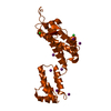
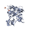
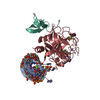
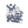
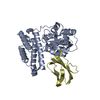
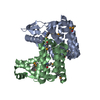
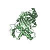
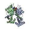
[English] 日本語
Yorodumi
- PDB-4xi7: Crystal structure of the MZM-REP domains of Mind bomb 1 in comple... -

+
Open data


ID or keywords:

Loading...

-
Basic information

Entry
Database: PDB / ID: 4xi7
TitleCrystal structure of the MZM-REP domains of Mind bomb 1 in complex with Jagged1 N-box peptide
Components
  • E3 ubiquitin-protein ligase MIB1
  • Jagged 1 N-box peptide
KeywordsLIGASE / E3 ligase / Notch signaling
Function / homology
Function and homology information


endocardial cushion cell development / loop of Henle development / ciliary body morphogenesis / regulation of reproductive process / pulmonary artery morphogenesis / cardiac neural crest cell development involved in outflow tract morphogenesis / podocyte development / negative regulation of endothelial cell differentiation / morphogenesis of an epithelial sheet / nephron development ...endocardial cushion cell development / loop of Henle development / ciliary body morphogenesis / regulation of reproductive process / pulmonary artery morphogenesis / cardiac neural crest cell development involved in outflow tract morphogenesis / podocyte development / negative regulation of endothelial cell differentiation / morphogenesis of an epithelial sheet / nephron development / positive regulation of myeloid cell differentiation / Nephron development / cardiac right ventricle morphogenesis / neural tube formation / inhibition of neuroepithelial cell differentiation / distal tubule development / neuroendocrine cell differentiation / aorta morphogenesis / Constitutive Signaling by NOTCH1 t(7;9)(NOTCH1:M1580_K2555) Translocation Mutant / positive regulation of cardiac epithelial to mesenchymal transition / NOTCH4 Activation and Transmission of Signal to the Nucleus / inner ear auditory receptor cell differentiation / T cell mediated immunity / endothelial cell differentiation / neuronal stem cell population maintenance / pulmonary valve morphogenesis / cell fate determination / negative regulation of stem cell differentiation / regulation of epithelial cell proliferation / aortic valve morphogenesis / Notch binding / myoblast differentiation / RUNX3 regulates NOTCH signaling / negative regulation of cell-matrix adhesion / negative regulation of cell-cell adhesion / positive regulation of Notch signaling pathway / negative regulation of fat cell differentiation / blood vessel development / heart looping / cardiac septum morphogenesis / positive regulation of endocytosis / response to muramyl dipeptide / hemopoiesis / centriolar satellite / negative regulation of neuron differentiation / RAC3 GTPase cycle / blood vessel remodeling / positive regulation of osteoblast differentiation / somitogenesis / keratinocyte differentiation / Notch signaling pathway / Constitutive Signaling by NOTCH1 HD Domain Mutants / NOTCH2 Activation and Transmission of Signal to the Nucleus / RAC1 GTPase cycle / Activated NOTCH1 Transmits Signal to the Nucleus / negative regulation of cell migration / NOTCH3 Activation and Transmission of Signal to the Nucleus / adherens junction / growth factor activity / RING-type E3 ubiquitin transferase / neuron differentiation / phospholipid binding / Constitutive Signaling by NOTCH1 PEST Domain Mutants / Constitutive Signaling by NOTCH1 HD+PEST Domain Mutants / endocytosis / ubiquitin-protein transferase activity / ubiquitin protein ligase activity / nervous system development / regulation of cell population proliferation / cytoplasmic vesicle / ubiquitin-dependent protein catabolic process / angiogenesis / in utero embryonic development / molecular adaptor activity / protein ubiquitination / apical plasma membrane / centrosome / calcium ion binding / structural molecule activity / positive regulation of transcription by RNA polymerase II / zinc ion binding / extracellular region / membrane / plasma membrane / cytosol
Similarity search - Function
Mind bomb, SH3 repeat domain / E3 ubiquitin-protein ligase MIB1/2, zinc finger, ZZ-type / Mind bomb SH3 repeat domain / Jagged/Serrate protein / Delta-like/Jagged, EGF-like domain / Delta/Serrate/lag-2 (DSL) protein / Notch ligand, N-terminal domain / Delta serrate ligand / N terminus of Notch ligand C2-like domain / DSL domain profile. ...Mind bomb, SH3 repeat domain / E3 ubiquitin-protein ligase MIB1/2, zinc finger, ZZ-type / Mind bomb SH3 repeat domain / Jagged/Serrate protein / Delta-like/Jagged, EGF-like domain / Delta/Serrate/lag-2 (DSL) protein / Notch ligand, N-terminal domain / Delta serrate ligand / N terminus of Notch ligand C2-like domain / DSL domain profile. / delta serrate ligand / Mib-herc2 / Mib/herc2 domain superfamily / Mib_herc2 / MIB/HERC2 domain profile. / EGF-like, conserved site / Human growth factor-like EGF / von Willebrand factor (vWF) type C domain / von Willebrand factor (vWF) type C domain / VWFC domain / : / Calcium-binding EGF domain / Zinc finger ZZ-type signature. / Zinc-binding domain, present in Dystrophin, CREB-binding protein. / Zinc finger, ZZ type / Zinc finger, ZZ-type / Zinc finger, ZZ-type superfamily / Zinc finger ZZ-type profile. / Ankyrin repeats (many copies) / Zinc finger, C3HC4 type (RING finger) / EGF-like domain / EGF-type aspartate/asparagine hydroxylation site / EGF-like calcium-binding, conserved site / Calcium-binding EGF-like domain signature. / Aspartic acid and asparagine hydroxylation site. / EGF-like calcium-binding domain / Calcium-binding EGF-like domain / Epidermal growth factor-like domain. / Ring finger / EGF-like domain profile. / Growth factor receptor cysteine-rich domain superfamily / EGF-like domain signature 2. / EGF-like domain signature 1. / EGF-like domain / Ankyrin repeats (3 copies) / Ankyrin repeat profile. / Ankyrin repeat region circular profile. / ankyrin repeats / Ankyrin repeat / Zinc finger RING-type profile. / Zinc finger, RING-type / Ankyrin repeat-containing domain superfamily / Zinc finger, RING/FYVE/PHD-type
Similarity search - Domain/homology
Protein jagged-1 / E3 ubiquitin-protein ligase MIB1
Similarity search - Component
Biological speciesHomo sapiens (human)
MethodX-RAY DIFFRACTION / SYNCHROTRON / MOLECULAR REPLACEMENT / Resolution: 2.051 Å
AuthorsMcMillan, B.J. / Blacklow, S.C.
Funding support United States, Germany, 3items
OrganizationGrant numberCountry
National Institutes of Health/National Cancer Institute (NIH/NCI)P01 CA119070 United States
American Cancer Society120246-PF-11-044-01-DMC United States
German Research Foundation (DFG)KL 1028/3-1 Germany
CitationJournal: Mol.Cell / Year: 2015
Title: A tail of two sites: a bipartite mechanism for recognition of notch ligands by mind bomb e3 ligases.
Authors: McMillan, B.J. / Schnute, B. / Ohlenhard, N. / Zimmerman, B. / Miles, L. / Beglova, N. / Klein, T. / Blacklow, S.C.
History
DepositionJan 6, 2015Deposition site: RCSB / Processing site: RCSB
Revision 1.0Mar 18, 2015Provider: repository / Type: Initial release
Revision 1.1Mar 25, 2015Group: Database references
Revision 1.2Sep 20, 2017Group: Author supporting evidence / Database references ...Author supporting evidence / Database references / Derived calculations / Refinement description / Source and taxonomy / Structure summary
Category: citation / entity_src_gen ...citation / entity_src_gen / pdbx_audit_support / pdbx_entity_src_syn / pdbx_struct_assembly / pdbx_struct_assembly_gen / pdbx_struct_assembly_prop / pdbx_struct_oper_list / software / struct_keywords
Item: _citation.journal_id_CSD / _entity_src_gen.pdbx_alt_source_flag ..._citation.journal_id_CSD / _entity_src_gen.pdbx_alt_source_flag / _pdbx_audit_support.funding_organization / _pdbx_entity_src_syn.pdbx_alt_source_flag / _pdbx_struct_assembly.oligomeric_details / _pdbx_struct_assembly_gen.asym_id_list / _pdbx_struct_assembly_prop.type / _pdbx_struct_assembly_prop.value / _pdbx_struct_oper_list.symmetry_operation / _software.classification / _struct_keywords.text
Revision 1.3Sep 27, 2017Group: Author supporting evidence / Category: pdbx_audit_support / Item: _pdbx_audit_support.funding_organization
Revision 1.4Dec 4, 2019Group: Author supporting evidence / Category: pdbx_audit_support / Item: _pdbx_audit_support.funding_organization
Revision 1.5Feb 28, 2024Group: Data collection / Database references / Category: chem_comp_atom / chem_comp_bond / database_2
Item: _database_2.pdbx_DOI / _database_2.pdbx_database_accession

-
Structure visualization

Structure viewerMolecule:
MolmilJmol/JSmol

Downloads & links

-
Assembly

Deposited unit
A: E3 ubiquitin-protein ligase MIB1
C: Jagged 1 N-box peptide
hetero molecules


Theoretical massNumber of molelcules
Total (without water)43,7526
Polymers43,4292
Non-polymers3234
Water2,900161
1


  • Idetical with deposited unit
  • defined by author&software
TypeNameSymmetry operationNumber
identity operation1_555x,y,z1
Buried area1040 Å2
ΔGint-27 kcal/mol
Surface area17600 Å2
MethodPISA
Unit cell
Length a, b, c (Å)73.728, 73.728, 170.276
Angle α, β, γ (deg.)90.000, 90.000, 90.000
Int Tables number91
Space group name H-MP4122

-
Components

#1: Protein E3 ubiquitin-protein ligase MIB1 / DAPK-interacting protein 1 / DIP-1 / Mind bomb homolog 1 / Zinc finger ZZ type with ankyrin repeat ...DAPK-interacting protein 1 / DIP-1 / Mind bomb homolog 1 / Zinc finger ZZ type with ankyrin repeat domain protein 2


Mass: 42148.145 Da / Num. of mol.: 1
Source method: isolated from a genetically manipulated source
Source: (gene. exp.) Homo sapiens (human) / Gene: MIB1, DIP1, KIAA1323, ZZANK2 / Production host: Escherichia coli (E. coli)
References: UniProt: Q86YT6, Ligases; Forming carbon-nitrogen bonds; Acid-amino-acid ligases (peptide synthases)
#2: Protein/peptide Jagged 1 N-box peptide


Mass: 1280.452 Da / Num. of mol.: 1 / Source method: obtained synthetically / Source: (synth.) Homo sapiens (human) / References: UniProt: P78504*PLUS
#3: Chemical ChemComp-ZN / ZINC ION


Mass: 65.409 Da / Num. of mol.: 2 / Source method: obtained synthetically / Formula: Zn
#4: Chemical ChemComp-SO4 / SULFATE ION


Mass: 96.063 Da / Num. of mol.: 2 / Source method: obtained synthetically / Formula: SO4
#5: Water ChemComp-HOH / water


Mass: 18.015 Da / Num. of mol.: 161 / Source method: isolated from a natural source / Formula: H2O

-
Experimental details

-
Experiment

ExperimentMethod: X-RAY DIFFRACTION / Number of used crystals: 1

-
Sample preparation

CrystalDensity Matthews: 2.66 Å3/Da / Density % sol: 53.77 %
Crystal growTemperature: 293.15 K / Method: vapor diffusion, hanging drop
Details: Protein at 6 mg/mL in 25 mM HEPES 7.4, 500 mM NaCl, 10% w/v glucose, 100 mM Arg, 2 mM TCEP Precipitant: 1.5 M (NH4)2SO4, 20% v/v glycerol, HEPES pH 7.6 1:1 drop volume

-
Data collection

DiffractionMean temperature: 100 K
Diffraction sourceSource: SYNCHROTRON / Site: APS / Beamline: 24-ID-E / Wavelength: 0.97918 Å
DetectorType: ADSC QUANTUM 315 / Detector: CCD / Date: Aug 8, 2013
RadiationProtocol: SINGLE WAVELENGTH / Monochromatic (M) / Laue (L): M / Scattering type: x-ray
Radiation wavelengthWavelength: 0.97918 Å / Relative weight: 1
ReflectionResolution: 2.05→50 Å / Num. obs: 29272 / % possible obs: 96.3 % / Redundancy: 10 % / Biso Wilson estimate: 29.36 Å2 / Rmerge(I) obs: 0.092 / Χ2: 1.079 / Net I/av σ(I): 26 / Net I/σ(I): 6.2 / Num. measured all: 293868
Reflection shell

Diffraction-ID: 1 / Rejects: _

Resolution (Å)Redundancy (%)Rmerge(I) obsNum. unique allΧ2% possible all
2.05-2.099.80.62714181.09594.5
2.09-2.1210.40.56914191.09197
2.12-2.1610.40.50714431.0997.1
2.16-2.2110.30.43614431.12297.1
2.21-2.2610.20.38914581.11397.5
2.26-2.3110.20.3414431.13697.3
2.31-2.3710.10.31414541.10197.1
2.37-2.439.80.26714351.09897
2.43-2.59.60.22213691.06791.5
2.5-2.5810.20.21714571.09496.9
2.58-2.6810.30.19614661.08497.9
2.68-2.7810.20.15914741.10497.8
2.78-2.9110.20.13214701.07597.7
2.91-3.06100.10615021.08397.2
3.06-3.259.70.07713771.04191.3
3.25-3.5110.20.06615051.08498
3.51-3.8610.20.05415161.01398.6
3.86-4.429.80.04515180.98497.4
4.42-5.569.90.04314741.03292.5
5.56-509.20.03916311.07594.8

-
Processing

Software
NameVersionClassification
PHENIXrefinement
PDB_EXTRACT3.15data extraction
HKL-2000data scaling
PHENIXphasing
Cootmodel building
RefinementMethod to determine structure: MOLECULAR REPLACEMENT / Resolution: 2.051→49.849 Å / SU ML: 0.2 / Cross valid method: FREE R-VALUE / σ(F): 1.36 / Phase error: 21.06 / Stereochemistry target values: ML
RfactorNum. reflection% reflection
Rfree0.2236 1480 5.06 %
Rwork0.1967 27746 -
obs0.198 29226 96.31 %
Solvent computationShrinkage radii: 0.9 Å / VDW probe radii: 1.11 Å / Solvent model: FLAT BULK SOLVENT MODEL
Displacement parametersBiso max: 153.24 Å2 / Biso mean: 44.892 Å2 / Biso min: 17.58 Å2
Refinement stepCycle: final / Resolution: 2.051→49.849 Å
ProteinNucleic acidLigandSolventTotal
Num. atoms2880 0 12 161 3053
Biso mean--32.87 35.72 -
Num. residues----368
Refine LS restraints
Refine-IDTypeDev idealNumber
X-RAY DIFFRACTIONf_bond_d0.0042961
X-RAY DIFFRACTIONf_angle_d0.7944001
X-RAY DIFFRACTIONf_chiral_restr0.032424
X-RAY DIFFRACTIONf_plane_restr0.004519
X-RAY DIFFRACTIONf_dihedral_angle_d12.1471063
LS refinement shell

Refine-ID: X-RAY DIFFRACTION / Total num. of bins used: 11

Resolution (Å)Rfactor RfreeNum. reflection RfreeRfactor RworkNum. reflection RworkNum. reflection all% reflection obs (%)
2.0506-2.11680.26181320.23252435256795
2.1168-2.19240.22681210.21832496261797
2.1924-2.28020.24941230.20782514263797
2.2802-2.3840.23361360.20482520265697
2.384-2.50970.27241080.20862425253393
2.5097-2.66690.23861560.21312510266698
2.6669-2.87280.24041400.21352533267398
2.8728-3.16180.25941390.20592526266597
3.1618-3.61920.20131380.19022515265396
3.6192-4.55940.17451470.1612608275598
4.5594-49.86420.24081400.20322664280493
Refinement TLS params.Method: refined / Origin x: -29.8341 Å / Origin y: -0.8355 Å / Origin z: -19.2426 Å
111213212223313233
T0.174 Å20.0093 Å20.0238 Å2-0.2474 Å20.0298 Å2--0.2125 Å2
L1.2163 °2-0.1835 °20.2367 °2-0.346 °2-0.2625 °2--0.8641 °2
S0.0252 Å °-0.1575 Å °0.0983 Å °0.0507 Å °0.0932 Å °0.0729 Å °-0.0423 Å °-0.2819 Å °-0.1159 Å °
Refinement TLS group
IDRefine-IDRefine TLS-IDSelection detailsAuth asym-IDAuth seq-ID
1X-RAY DIFFRACTION1allA7 - 400
2X-RAY DIFFRACTION1allB1 - 2
3X-RAY DIFFRACTION1allC2 - 7
4X-RAY DIFFRACTION1allD1 - 2
5X-RAY DIFFRACTION1allE1 - 267

+
About Yorodumi

-
News

-
Feb 9, 2022. New format data for meta-information of EMDB entries

New format data for meta-information of EMDB entries

  • Version 3 of the EMDB header file is now the official format.
  • The previous official version 1.9 will be removed from the archive.

Related info.:EMDB header

External links:wwPDB to switch to version 3 of the EMDB data model

-
Aug 12, 2020. Covid-19 info

Covid-19 info

URL: https://pdbjlvh1.pdbj.org/emnavi/covid19.php

New page: Covid-19 featured information page in EM Navigator.

Related info.:Covid-19 info / Mar 5, 2020. Novel coronavirus structure data

+
Mar 5, 2020. Novel coronavirus structure data

Novel coronavirus structure data

Related info.:Yorodumi Speices / Aug 12, 2020. Covid-19 info

External links:COVID-19 featured content - PDBj / Molecule of the Month (242):Coronavirus Proteases

+
Jan 31, 2019. EMDB accession codes are about to change! (news from PDBe EMDB page)

EMDB accession codes are about to change! (news from PDBe EMDB page)

  • The allocation of 4 digits for EMDB accession codes will soon come to an end. Whilst these codes will remain in use, new EMDB accession codes will include an additional digit and will expand incrementally as the available range of codes is exhausted. The current 4-digit format prefixed with “EMD-” (i.e. EMD-XXXX) will advance to a 5-digit format (i.e. EMD-XXXXX), and so on. It is currently estimated that the 4-digit codes will be depleted around Spring 2019, at which point the 5-digit format will come into force.
  • The EM Navigator/Yorodumi systems omit the EMD- prefix.

Related info.:Q: What is EMD? / ID/Accession-code notation in Yorodumi/EM Navigator

External links:EMDB Accession Codes are Changing Soon! / Contact to PDBj

+
Jul 12, 2017. Major update of PDB

Major update of PDB

  • wwPDB released updated PDB data conforming to the new PDBx/mmCIF dictionary.
  • This is a major update changing the version number from 4 to 5, and with Remediation, in which all the entries are updated.
  • In this update, many items about electron microscopy experimental information are reorganized (e.g. em_software).
  • Now, EM Navigator and Yorodumi are based on the updated data.

External links:wwPDB Remediation / Enriched Model Files Conforming to OneDep Data Standards Now Available in the PDB FTP Archive

-
Yorodumi

Thousand views of thousand structures

  • Yorodumi is a browser for structure data from EMDB, PDB, SASBDB, etc.
  • This page is also the successor to EM Navigator detail page, and also detail information page/front-end page for Omokage search.
  • The word "yorodu" (or yorozu) is an old Japanese word meaning "ten thousand". "mi" (miru) is to see.

Related info.:EMDB / PDB / SASBDB / Comparison of 3 databanks / Yorodumi Search / Aug 31, 2016. New EM Navigator & Yorodumi / Yorodumi Papers / Jmol/JSmol / Function and homology information / Changes in new EM Navigator and Yorodumi

Read more