 Movie
Movie Controller


+ Open data

- Basic information
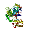
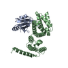
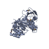
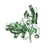
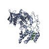
| Entry | Database: PDB / ID: 3eb8 | ||||||
|---|---|---|---|---|---|---|---|
| Title | VirA | ||||||
 Components Components | Cysteine protease-like virA | ||||||
 Keywords Keywords | HYDROLASE / beta sheet / alpha helix / Protease / Secreted / Thiol protease / Virulence | ||||||
| Function / homology |  Function and homology information Function and homology informationHydrolases; Acting on peptide bonds (peptidases); Cysteine endopeptidases / cysteine-type endopeptidase activity / proteolysis / extracellular region Similarity search - Function | ||||||
| Biological species |  Shigella flexneri (bacteria) Shigella flexneri (bacteria) | ||||||
| Method |  X-RAY DIFFRACTION / SYNCHROTRON / SIRAS / Resolution: 2.4 Å X-RAY DIFFRACTION / SYNCHROTRON / SIRAS / Resolution: 2.4 Å | ||||||
 Authors Authors | Germane, K.L. / Spiller, B.W. | ||||||
 Citation Citation | Journal: Biochemistry / Year: 2008 Title: Structural and functional studies indicate that Shigella VirA is not a protease and does not directly destabilize microtubules. Authors: Germane, K.L. / Ohi, R. / Goldberg, M.B. / Spiller, B.W. | ||||||
| History |
|
- Structure visualization
| Structure viewer | Molecule: MolmilJmol/JSmol |
|---|
- Downloads & links
-Download
| PDBx/mmCIF format | 3eb8.cif.gz | 272.3 KB | Display | PDBx/mmCIF format |
|---|---|---|---|---|
| PDB format | pdb3eb8.ent.gz | 223.7 KB | Display | PDB format |
| PDBx/mmJSON format | 3eb8.json.gz | Tree view | PDBx/mmJSON format | |
| Others |  Other downloads Other downloads |
-Validation report
| Arichive directory | https://data.pdbj.org/pub/pdb/validation_reports/eb/3eb8ftp://data.pdbj.org/pub/pdb/validation_reports/eb/3eb8 | HTTPS FTP |
|---|
-Related structure data
| Similar structure data |
|---|


-Links
 PDBj
PDBj- Assembly
| Deposited unit | 
| ||||||||
|---|---|---|---|---|---|---|---|---|---|
| 1 | 
| ||||||||
| 2 | 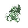
| ||||||||
| 3 | 
| ||||||||
| Unit cell |
|
-Components
| #1: Protein | Mass: 39872.914 Da / Num. of mol.: 2 / Fragment: delta44 Source method: isolated from a genetically manipulated source Source: (gene. exp.) Shigella flexneri (bacteria) / Gene: virA, CP0181, pWR501_0191 / Plasmid: pET28a / Production host: References: UniProt: Q7BU69, Hydrolases; Acting on peptide bonds (peptidases); Cysteine endopeptidases #2: Water | ChemComp-HOH / |  Mass: 18.015 Da / Num. of mol.: 91 / Source method: isolated from a natural source / Formula: H2O Mass: 18.015 Da / Num. of mol.: 91 / Source method: isolated from a natural source / Formula: H2O |
|---|
-Experimental details
-Experiment
| Experiment | Method: X-RAY DIFFRACTION |
|---|
- Sample preparation
| Crystal | Density Matthews: 2.59 Å3/Da / Density % sol: 52.51 % |
|---|
-Data collection
| Diffraction | Mean temperature: 100 K |
|---|---|
| Diffraction source | Source: SYNCHROTRON / Site: APS  / Beamline: 24-ID-C / Wavelength: 0.97919 / Beamline: 24-ID-C / Wavelength: 0.97919 |
| Detector | Type: ADSC QUANTUM 4 / Detector: CCD |
| Radiation | Protocol: SINGLE WAVELENGTH / Monochromatic (M) / Laue (L): M / Scattering type: x-ray |
| Radiation wavelength | Wavelength: 0.97919 Å / Relative weight: 1 |
| Reflection | Resolution: 2.4→40 Å / Num. obs: 33386 / % possible obs: 99.5 % / Observed criterion σ(I): 2 / Redundancy: 6.3 % / Biso Wilson estimate: 59.8 Å2 / Rmerge(I) obs: 0.054 / Net I/σ(I): 27 |
| Reflection shell | Resolution: 2.4→2.49 Å / Redundancy: 5 % / Rmerge(I) obs: 0.386 / Mean I/σ(I) obs: 2.07 / % possible all: 98 |
- Processing
| Software |
| |||||||||||||||||||||||||||||||||||||||||||||||||||||||||||||||||||||||||||||||||||||||||||
|---|---|---|---|---|---|---|---|---|---|---|---|---|---|---|---|---|---|---|---|---|---|---|---|---|---|---|---|---|---|---|---|---|---|---|---|---|---|---|---|---|---|---|---|---|---|---|---|---|---|---|---|---|---|---|---|---|---|---|---|---|---|---|---|---|---|---|---|---|---|---|---|---|---|---|---|---|---|---|---|---|---|---|---|---|---|---|---|---|---|---|---|---|
| Refinement | Method to determine structure: SIRAS / Resolution: 2.4→39.99 Å / SU ML: 0.39 / Stereochemistry target values: ML
| |||||||||||||||||||||||||||||||||||||||||||||||||||||||||||||||||||||||||||||||||||||||||||
| Solvent computation | Shrinkage radii: 0.9 Å / VDW probe radii: 1.11 Å / Solvent model: FLAT BULK SOLVENT MODEL / Bsol: 58.18 Å2 / ksol: 0.34 e/Å3 | |||||||||||||||||||||||||||||||||||||||||||||||||||||||||||||||||||||||||||||||||||||||||||
| Displacement parameters | Biso mean: 62.43 Å2
| |||||||||||||||||||||||||||||||||||||||||||||||||||||||||||||||||||||||||||||||||||||||||||
| Refinement step | Cycle: LAST / Resolution: 2.4→39.99 Å
| |||||||||||||||||||||||||||||||||||||||||||||||||||||||||||||||||||||||||||||||||||||||||||
| LS refinement shell |
|
