 Movie
Movie Controller


+ Open data

- Basic information
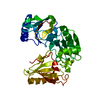
| Entry | Database: PDB / ID: 1p8x | ||||||
|---|---|---|---|---|---|---|---|
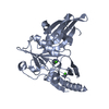
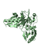
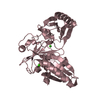
| Title | The Calcium-Activated C-terminal half of gelsolin | ||||||
 Components Components | Gelsolin precursor, plasma | ||||||
 Keywords Keywords | STRUCTURAL PROTEIN / calcium-binding | ||||||
| Function / homology |  Function and homology information Function and homology informationstriated muscle atrophy / regulation of establishment of T cell polarity / regulation of plasma membrane raft polarization / regulation of receptor clustering / positive regulation of keratinocyte apoptotic process / renal protein absorption / positive regulation of protein processing in phagocytic vesicle / phosphatidylinositol 3-kinase catalytic subunit binding / positive regulation of actin nucleation / actin cap ...striated muscle atrophy / regulation of establishment of T cell polarity / regulation of plasma membrane raft polarization / regulation of receptor clustering / positive regulation of keratinocyte apoptotic process / renal protein absorption / positive regulation of protein processing in phagocytic vesicle / phosphatidylinositol 3-kinase catalytic subunit binding / positive regulation of actin nucleation / actin cap / myosin II binding / host-mediated suppression of symbiont invasion / cell projection assembly / actin filament severing / barbed-end actin filament capping / actin polymerization or depolymerization / actin filament depolymerization / actin filament capping / relaxation of cardiac muscle / Sensory processing of sound by outer hair cells of the cochlea / phagocytosis, engulfment / cardiac muscle cell contraction / hepatocyte apoptotic process / sarcoplasm / cilium assembly / Caspase-mediated cleavage of cytoskeletal proteins / phagocytic vesicle / response to muscle stretch / actin filament polymerization / phosphatidylinositol-4,5-bisphosphate binding / actin filament organization / central nervous system development / cellular response to type II interferon / protein destabilization / actin filament binding / lamellipodium / actin cytoskeleton / actin binding / secretory granule lumen / blood microparticle / amyloid fibril formation / ficolin-1-rich granule lumen / Amyloid fiber formation / focal adhesion / calcium ion binding / positive regulation of gene expression / Neutrophil degranulation / : / extracellular exosome / extracellular region / plasma membrane / cytosol / cytoplasm Similarity search - Function | ||||||
| Biological species |  Homo sapiens (human) Homo sapiens (human) | ||||||
| Method |  X-RAY DIFFRACTION / SYNCHROTRON / MOLECULAR REPLACEMENT / Resolution: 2 Å X-RAY DIFFRACTION / SYNCHROTRON / MOLECULAR REPLACEMENT / Resolution: 2 Å | ||||||
 Authors Authors | Narayan, K. / Burtnick, L.D. / Robinson, R.C. | ||||||
 Citation Citation | Journal: FEBS LETT. / Year: 2003 Title: Activation in isolation: Exposure of the actin-binding site in the C-terminal half of gelsolin does not require actin Authors: Narayan, K. / Chumnarnsilpa, S. / Choe, H. / Irobi, E. / Urosev, D. / Lindberg, U. / Schutt, C.E. / Burtnick, L.D. / Robinson, R.C. | ||||||
| History |
|
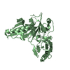
- Structure visualization
| Structure viewer | Molecule: MolmilJmol/JSmol |
|---|
- Downloads & links
-Download
| PDBx/mmCIF format | 1p8x.cif.gz | 223.6 KB | Display | PDBx/mmCIF format |
|---|---|---|---|---|
| PDB format | pdb1p8x.ent.gz | 177.1 KB | Display | PDB format |
| PDBx/mmJSON format | 1p8x.json.gz | Tree view | PDBx/mmJSON format | |
| Others |  Other downloads Other downloads |
-Validation report
| Arichive directory | https://data.pdbj.org/pub/pdb/validation_reports/p8/1p8xftp://data.pdbj.org/pub/pdb/validation_reports/p8/1p8x | HTTPS FTP |
|---|
-Related structure data
| Related structure data | 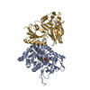 1h1vS S: Starting model for refinement |
|---|---|
| Similar structure data |


-Links
 PDBj
PDBj

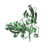
- Assembly
| Deposited unit | 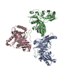
| ||||||||
|---|---|---|---|---|---|---|---|---|---|
| 1 | 
| ||||||||
| 2 | 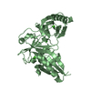
| ||||||||
| 3 | 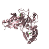
| ||||||||
| Unit cell |
|
-Components
| #1: Protein | Mass: 37762.098 Da / Num. of mol.: 3 / Fragment: C-terminal domain Source method: isolated from a genetically manipulated source Source: (gene. exp.) Homo sapiens (human) / Plasmid: pGEX-6P-1 / Production host:  #2: Chemical | ChemComp-CA /   Mass: 40.078 Da / Num. of mol.: 9 / Source method: obtained synthetically / Formula: Ca Mass: 40.078 Da / Num. of mol.: 9 / Source method: obtained synthetically / Formula: Ca#3: Water | ChemComp-HOH / |  Mass: 18.015 Da / Num. of mol.: 1040 / Source method: isolated from a natural source / Formula: H2O Mass: 18.015 Da / Num. of mol.: 1040 / Source method: isolated from a natural source / Formula: H2O |
|---|
-Experimental details
-Experiment
| Experiment | Method: X-RAY DIFFRACTION / Number of used crystals: 1 |
|---|
- Sample preparation
| Crystal | Density Matthews: 2.64 Å3/Da / Density % sol: 53.47 % | ||||||||||||||||||||||||
|---|---|---|---|---|---|---|---|---|---|---|---|---|---|---|---|---|---|---|---|---|---|---|---|---|---|
| Crystal grow | Temperature: 277.15 K / Method: vapor diffusion, hanging drop / pH: 7.5 Details: 15% (w/v) PEG 8000, 100 mM Tris-HCl, pH 7.5, VAPOR DIFFUSION, HANGING DROP, temperature 277.15K | ||||||||||||||||||||||||
| Crystal grow | *PLUS Temperature: 4 ℃ / Method: vapor diffusion, hanging drop | ||||||||||||||||||||||||
| Components of the solutions | *PLUS
|
-Data collection
| Diffraction | Mean temperature: 100 K |
|---|---|
| Diffraction source | Source: SYNCHROTRON / Site: ESRF  / Beamline: ID14-4 / Wavelength: 1.068 Å / Beamline: ID14-4 / Wavelength: 1.068 Å |
| Detector | Type: ADSC QUANTUM 4 / Detector: CCD / Date: Apr 13, 2003 |
| Radiation | Protocol: SINGLE WAVELENGTH / Monochromatic (M) / Laue (L): M / Scattering type: x-ray |
| Radiation wavelength | Wavelength: 1.068 Å / Relative weight: 1 |
| Reflection | Resolution: 2→20 Å / % possible obs: 99.2 % / Observed criterion σ(F): 0 / Observed criterion σ(I): 0 / Redundancy: 6.3 % / Biso Wilson estimate: 31 Å2 / Rmerge(I) obs: 0.054 / Net I/σ(I): 6.6 |
| Reflection shell | Resolution: 2→2.1 Å / Redundancy: 6.1 % / Rmerge(I) obs: 0.13 / Mean I/σ(I) obs: 6 / Num. unique all: 11619 / % possible all: 98.7 |
| Reflection | *PLUS Highest resolution: 2 Å / Lowest resolution: 20 Å / Num. obs: 81129 / Num. measured all: 512059 |
| Reflection shell | *PLUS % possible obs: 98.7 % / Num. unique obs: 11619 / Num. measured obs: 70689 / Rmerge(I) obs: 0.13 |
- Processing
| Software |
| ||||||||||||||||||||||||||||
|---|---|---|---|---|---|---|---|---|---|---|---|---|---|---|---|---|---|---|---|---|---|---|---|---|---|---|---|---|---|
| Refinement | Method to determine structure: MOLECULAR REPLACEMENT Starting model: The gelsolin (G4-G6) portion of PDB Entry 1H1V Resolution: 2→20 Å / Cross valid method: THROUGHOUT / σ(F): 0 / Stereochemistry target values: Engh & Huber
| ||||||||||||||||||||||||||||
| Displacement parameters |
| ||||||||||||||||||||||||||||
| Refinement step | Cycle: LAST / Resolution: 2→20 Å
| ||||||||||||||||||||||||||||
| Refine LS restraints |
| ||||||||||||||||||||||||||||
| LS refinement shell | Resolution: 2→2.1 Å
| ||||||||||||||||||||||||||||
| Xplor file |
| ||||||||||||||||||||||||||||
| Refinement | *PLUS % reflection Rfree: 5 % | ||||||||||||||||||||||||||||
| Solvent computation | *PLUS | ||||||||||||||||||||||||||||
| Displacement parameters | *PLUS |
