 Movie
Movie Controller

[English] 日本語
 Yorodumi
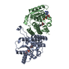
- PDB-4ww5: Crystal structure of binary complex Bud32-Cgi121 in complex with AMPP -
+ Open data

- Basic information
| Entry | Database: PDB / ID: 4ww5 | ||||||
|---|---|---|---|---|---|---|---|
| Title | Crystal structure of binary complex Bud32-Cgi121 in complex with AMPP | ||||||
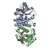
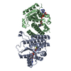
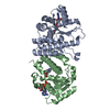
 Components Components | (EKC/KEOPS complex subunit ...) x 2 | ||||||
 Keywords Keywords | TRANSFERASE / KEOPS / binary complex / Bud32-Cgi121 / tRNA t6A | ||||||
| Function / homology |  Function and homology information Function and homology informationEKC/KEOPS complex / tRNA threonylcarbamoyladenosine metabolic process / cellular bud site selection / tRNA threonylcarbamoyladenosine modification / Hydrolases; Acting on acid anhydrides / telomere maintenance via recombination / telomere maintenance / maintenance of translational fidelity / DNA recombination / chromosome, telomeric region ...EKC/KEOPS complex / tRNA threonylcarbamoyladenosine metabolic process / cellular bud site selection / tRNA threonylcarbamoyladenosine modification / Hydrolases; Acting on acid anhydrides / telomere maintenance via recombination / telomere maintenance / maintenance of translational fidelity / DNA recombination / chromosome, telomeric region / non-specific serine/threonine protein kinase / tRNA binding / protein serine kinase activity / protein serine/threonine kinase activity / positive regulation of transcription by RNA polymerase II / ATP hydrolysis activity / nucleoplasm / ATP binding / nucleus / cytosol / cytoplasm Similarity search - Function | ||||||
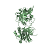
| Biological species |  | ||||||
| Method |  X-RAY DIFFRACTION / SYNCHROTRON / MOLECULAR REPLACEMENT / Resolution: 1.997 Å X-RAY DIFFRACTION / SYNCHROTRON / MOLECULAR REPLACEMENT / Resolution: 1.997 Å | ||||||
 Authors Authors | Zhang, W. | ||||||
 Citation Citation | Journal: Nucleic Acids Res. / Year: 2015 Title: Crystal structures of the Gon7/Pcc1 and Bud32/Cgi121 complexes provide a model for the complete yeast KEOPS complex. Authors: Zhang, W. / Collinet, B. / Graille, M. / Daugeron, M.C. / Lazar, N. / Libri, D. / Durand, D. / van Tilbeurgh, H. | ||||||
| History |
|
- Structure visualization
| Structure viewer | Molecule: MolmilJmol/JSmol |
|---|
- Downloads & links
-Download
| PDBx/mmCIF format | 4ww5.cif.gz | 102.4 KB | Display | PDBx/mmCIF format |
|---|---|---|---|---|
| PDB format | pdb4ww5.ent.gz | 75.3 KB | Display | PDB format |
| PDBx/mmJSON format | 4ww5.json.gz | Tree view | PDBx/mmJSON format | |
| Others |  Other downloads Other downloads |
-Validation report
| Arichive directory | https://data.pdbj.org/pub/pdb/validation_reports/ww/4ww5ftp://data.pdbj.org/pub/pdb/validation_reports/ww/4ww5 | HTTPS FTP |
|---|
-Related structure data
| Related structure data | 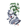 4ww7C 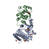 4ww9C 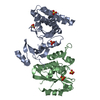 4wwaC 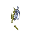 4wx8C 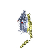 4wxaC  4xahC C: citing same article ( |
|---|---|
| Similar structure data |


-Links
 PDBj
PDBj


- Assembly
| Deposited unit | 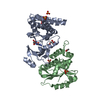
| ||||||||
|---|---|---|---|---|---|---|---|---|---|
| 1 |
| ||||||||
| Unit cell |
|
-Components
-EKC/KEOPS complex subunit ... , 2 types, 2 molecules AB
| #1: Protein | Mass: 29982.377 Da / Num. of mol.: 1 Source method: isolated from a genetically manipulated source Source: (gene. exp.) Strain: ATCC 204508 / S288c / Gene: BUD32, LDB14, YGR262C Production host:  References: UniProt: P53323, Hydrolases; Acting on acid anhydrides, non-specific serine/threonine protein kinase |
|---|---|
| #2: Protein | Mass: 20686.812 Da / Num. of mol.: 1 Source method: isolated from a genetically manipulated source Source: (gene. exp.) Strain: ATCC 204508 / S288c / Gene: CGI121, YML036W Production host: References: UniProt: Q03705 |
-Non-polymers , 5 types, 176 molecules 


| #3: Chemical | ChemComp-ANP /  Mass: 506.196 Da / Num. of mol.: 1 / Source method: obtained synthetically / Formula: C10H17N6O12P3 / Comment: AMP-PNP, energy-carrying molecule analogue*YM Mass: 506.196 Da / Num. of mol.: 1 / Source method: obtained synthetically / Formula: C10H17N6O12P3 / Comment: AMP-PNP, energy-carrying molecule analogue*YM | ||||||
|---|---|---|---|---|---|---|---|
| #4: Chemical |  Mass: 59.044 Da / Num. of mol.: 3 / Source method: obtained synthetically / Formula: C2H3O2 Mass: 59.044 Da / Num. of mol.: 3 / Source method: obtained synthetically / Formula: C2H3O2#5: Chemical | ChemComp-SO4 /  Mass: 96.063 Da / Num. of mol.: 5 / Source method: obtained synthetically / Formula: SO4 Mass: 96.063 Da / Num. of mol.: 5 / Source method: obtained synthetically / Formula: SO4#6: Chemical | ChemComp-GOL / |  Mass: 92.094 Da / Num. of mol.: 1 / Source method: obtained synthetically / Formula: C3H8O3 Mass: 92.094 Da / Num. of mol.: 1 / Source method: obtained synthetically / Formula: C3H8O3#7: Water | ChemComp-HOH / | Mass: 18.015 Da / Num. of mol.: 166 / Source method: isolated from a natural source / Formula: H2O |
-Experimental details
-Experiment
| Experiment | Method: X-RAY DIFFRACTION |
|---|
- Sample preparation
| Crystal | Density Matthews: 2.74 Å3/Da / Density % sol: 55.18 % |
|---|---|
| Crystal grow | Temperature: 293 K / Method: vapor diffusion, hanging drop / pH: 4.6 Details: 0.1 M Sodium Acetate pH 4.6, 2.0 M Ammounium Sulfate, 0.1 M NaCl,10 mM Tris-HCl pH 7.5, 5 mM AMPPNP and 5 mM L-threonine. PH range: 4.6-5.3 |
-Data collection
| Diffraction | Mean temperature: 100 K |
|---|---|
| Diffraction source | Source: SYNCHROTRON / Site: SOLEIL  / Beamline: PROXIMA 1 / Wavelength: 0.98011 Å / Beamline: PROXIMA 1 / Wavelength: 0.98011 Å |
| Detector | Type: PSI PILATUS 6M / Detector: PIXEL / Date: Dec 4, 2011 |
| Radiation | Protocol: SINGLE WAVELENGTH / Monochromatic (M) / Laue (L): M / Scattering type: x-ray |
| Radiation wavelength | Wavelength: 0.98011 Å / Relative weight: 1 |
| Reflection | Resolution: 1.997→39.93 Å / Num. all: 283335 / Num. obs: 37119 / % possible obs: 99.79 % / Redundancy: 7.6 % / Rsym value: 0.049 / Net I/σ(I): 27.29 |
| Reflection shell | Resolution: 1.997→2.069 Å / Mean I/σ(I) obs: 5.54 / % possible all: 98.1 |
- Processing
| Software | Name: PHENIX / Version: (phenix.refine) / Classification: refinement | ||||||||||||||||||||||||||||||||||||||||||||||||||||||||||||||||||||||||||||||||||||||||||||||||||
|---|---|---|---|---|---|---|---|---|---|---|---|---|---|---|---|---|---|---|---|---|---|---|---|---|---|---|---|---|---|---|---|---|---|---|---|---|---|---|---|---|---|---|---|---|---|---|---|---|---|---|---|---|---|---|---|---|---|---|---|---|---|---|---|---|---|---|---|---|---|---|---|---|---|---|---|---|---|---|---|---|---|---|---|---|---|---|---|---|---|---|---|---|---|---|---|---|---|---|---|
| Refinement | Method to determine structure: MOLECULAR REPLACEMENT / Resolution: 1.997→39.927 Å / SU ML: 0.27 / Cross valid method: THROUGHOUT / σ(F): 1.99 / Phase error: 21.77 / Stereochemistry target values: ML
| ||||||||||||||||||||||||||||||||||||||||||||||||||||||||||||||||||||||||||||||||||||||||||||||||||
| Solvent computation | Shrinkage radii: 0.9 Å / VDW probe radii: 1.11 Å / Solvent model: FLAT BULK SOLVENT MODEL / Bsol: 50.862 Å2 / ksol: 0.386 e/Å3 | ||||||||||||||||||||||||||||||||||||||||||||||||||||||||||||||||||||||||||||||||||||||||||||||||||
| Displacement parameters |
| ||||||||||||||||||||||||||||||||||||||||||||||||||||||||||||||||||||||||||||||||||||||||||||||||||
| Refinement step | Cycle: LAST / Resolution: 1.997→39.927 Å
| ||||||||||||||||||||||||||||||||||||||||||||||||||||||||||||||||||||||||||||||||||||||||||||||||||
| Refine LS restraints |
| ||||||||||||||||||||||||||||||||||||||||||||||||||||||||||||||||||||||||||||||||||||||||||||||||||
| LS refinement shell |
|
