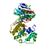
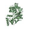
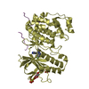
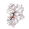
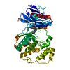
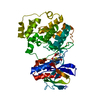
[English] 日本語
Yorodumi
- PDB-4wua: Crystal structure of human SRPK1 complexed to an inhibitor SRPIN340 -

+
Open data


ID or keywords:

Loading...

-
Basic information

Entry
Database: PDB / ID: 4wua
TitleCrystal structure of human SRPK1 complexed to an inhibitor SRPIN340
ComponentsSRSF protein kinase 1, linker, SRSF protein kinase 1
KeywordsTRANSFERASE/TRANSFERASE INHIBITOR / protein kinase / kinase-inhibitor complex / pre-mRNA splicing / ATP-binding / TRANSFERASE-TRANSFERASE INHIBITOR complex
Function / homology
Function and homology information


sperm DNA condensation / regulation of mRNA processing / regulation of mRNA splicing, via spliceosome / Maturation of nucleoprotein / negative regulation of viral genome replication / spliceosomal complex assembly / positive regulation of viral genome replication / Replacement of protamines by nucleosomes in the male pronucleus / RNA splicing / chromosome segregation ...sperm DNA condensation / regulation of mRNA processing / regulation of mRNA splicing, via spliceosome / Maturation of nucleoprotein / negative regulation of viral genome replication / spliceosomal complex assembly / positive regulation of viral genome replication / Replacement of protamines by nucleosomes in the male pronucleus / RNA splicing / chromosome segregation / nuclear matrix / eukaryotic translation initiation factor 2alpha kinase activity / 3-phosphoinositide-dependent protein kinase activity / DNA-dependent protein kinase activity / ribosomal protein S6 kinase activity / histone H3S10 kinase activity / histone H2AXS139 kinase activity / histone H3S28 kinase activity / histone H4S1 kinase activity / histone H2BS14 kinase activity / histone H3T3 kinase activity / histone H2AS121 kinase activity / Rho-dependent protein serine/threonine kinase activity / histone H2BS36 kinase activity / histone H3S57 kinase activity / histone H2AT120 kinase activity / AMP-activated protein kinase activity / histone H2AS1 kinase activity / histone H3T6 kinase activity / histone H3T11 kinase activity / histone H3T45 kinase activity / non-specific serine/threonine protein kinase / protein kinase activity / intracellular signal transduction / nuclear speck / protein phosphorylation / innate immune response / protein serine kinase activity / protein serine/threonine kinase activity / chromatin / magnesium ion binding / endoplasmic reticulum / RNA binding / nucleoplasm / ATP binding / nucleus / plasma membrane / cytosol / cytoplasm
Similarity search - Function
: / Phosphorylase Kinase; domain 1 / Phosphorylase Kinase; domain 1 / Transferase(Phosphotransferase) domain 1 / Transferase(Phosphotransferase); domain 1 / Serine/threonine-protein kinase, active site / Serine/Threonine protein kinases active-site signature. / Protein kinase domain / Serine/Threonine protein kinases, catalytic domain / Protein kinase, ATP binding site ...: / Phosphorylase Kinase; domain 1 / Phosphorylase Kinase; domain 1 / Transferase(Phosphotransferase) domain 1 / Transferase(Phosphotransferase); domain 1 / Serine/threonine-protein kinase, active site / Serine/Threonine protein kinases active-site signature. / Protein kinase domain / Serine/Threonine protein kinases, catalytic domain / Protein kinase, ATP binding site / Protein kinases ATP-binding region signature. / Protein kinase domain profile. / Protein kinase domain / Protein kinase-like domain superfamily / 2-Layer Sandwich / Orthogonal Bundle / Mainly Alpha / Alpha Beta
Similarity search - Domain/homology
Chem-3UL / CITRIC ACID / SRSF protein kinase 1
Similarity search - Component
Biological speciesHomo sapiens (human)
MethodX-RAY DIFFRACTION / SYNCHROTRON / Resolution: 2 Å
AuthorsHoshina, M. / Ikura, T. / Hosoya, T. / Hagiwara, M. / Ito, N.
CitationJournal: Mol.Pharmacol. / Year: 2015
Title: Identification of a Dual Inhibitor of SRPK1 and CK2 That Attenuates Pathological Angiogenesis of Macular Degeneration in Mice
Authors: Morooka, S. / Hoshina, M. / Kii, I. / Okabe, T. / Kojima, H. / Inoue, N. / Okuno, Y. / Denawa, M. / Yoshida, S. / Fukuhara, J. / Ninomiya, K. / Ikura, T. / Furuya, T. / Nagano, T. / Noda, K. ...Authors: Morooka, S. / Hoshina, M. / Kii, I. / Okabe, T. / Kojima, H. / Inoue, N. / Okuno, Y. / Denawa, M. / Yoshida, S. / Fukuhara, J. / Ninomiya, K. / Ikura, T. / Furuya, T. / Nagano, T. / Noda, K. / Ishida, S. / Hosoya, T. / Ito, N. / Yoshimura, N. / Hagiwara, M.
History
DepositionOct 31, 2014Deposition site: RCSB / Processing site: PDBJ
Revision 1.0Sep 16, 2015Provider: repository / Type: Initial release
Revision 1.1Feb 5, 2020Group: Data collection / Database references / Derived calculations
Category: citation / diffrn_source ...citation / diffrn_source / pdbx_prerelease_seq / pdbx_struct_oper_list
Item: _citation.journal_id_CSD / _diffrn_source.pdbx_synchrotron_site / _pdbx_struct_oper_list.symmetry_operation
Revision 1.2Nov 8, 2023Group: Data collection / Database references / Refinement description
Category: chem_comp_atom / chem_comp_bond ...chem_comp_atom / chem_comp_bond / database_2 / pdbx_initial_refinement_model
Item: _database_2.pdbx_DOI / _database_2.pdbx_database_accession

-
Structure visualization

Structure viewerMolecule:
MolmilJmol/JSmol

Downloads & links

-
Assembly

Deposited unit
A: SRSF protein kinase 1, linker, SRSF protein kinase 1
hetero molecules


Theoretical massNumber of molelcules
Total (without water)46,1813
Polymers45,6391
Non-polymers5412
Water2,036113
1


  • Idetical with deposited unit
  • defined by author&software
TypeNameSymmetry operationNumber
identity operation1_555x,y,z1
Buried area180 Å2
ΔGint1 kcal/mol
Surface area16450 Å2
MethodPISA
Unit cell
Length a, b, c (Å)75.132, 75.132, 310.623
Angle α, β, γ (deg.)90.00, 90.00, 120.00
Int Tables number179
Space group name H-MP6522

-
Components

#1: Protein SRSF protein kinase 1, linker, SRSF protein kinase 1 / SFRS protein kinase 1 / Serine/arginine-rich protein-specific kinase 1 / SR-protein-specific kinase 1


Mass: 45639.066 Da / Num. of mol.: 1 / Fragment: UNP residues 213-426, UNP residues 465-826
Source method: isolated from a genetically manipulated source
Source: (gene. exp.) Homo sapiens (human) / Gene: SRPK1 / Production host: Escherichia coli (E. coli) / Strain (production host): BL-21(DE3)
References: UniProt: Q96SB4, non-specific serine/threonine protein kinase
#2: Chemical ChemComp-3UL / N-[2-(1-piperidinyl)-5-(trifluoromethyl)phenyl]-4-pyridinecarboxamide


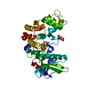
Mass: 349.350 Da / Num. of mol.: 1 / Source method: obtained synthetically / Formula: C18H18F3N3O
#3: Chemical ChemComp-CIT / CITRIC ACID


Mass: 192.124 Da / Num. of mol.: 1 / Source method: obtained synthetically / Formula: C6H8O7
#4: Water ChemComp-HOH / water


Mass: 18.015 Da / Num. of mol.: 113 / Source method: isolated from a natural source / Formula: H2O

-
Experimental details

-
Experiment

ExperimentMethod: X-RAY DIFFRACTION

-
Sample preparation

CrystalDensity Matthews: 2.77 Å3/Da / Density % sol: 55.64 %
Description: THE ENTRY CONTAINS FRIEDEL PAIRS IN F_PLUS/MINUS COLUMNS.
Crystal growTemperature: 293 K / Method: vapor diffusion, hanging drop / pH: 5.6
Details: 0.1M sodium citrate, 0.2M ammonium acetate, 5%PEG6000

-
Data collection

DiffractionMean temperature: 100 K
Diffraction sourceSource: SYNCHROTRON / Site: SPring-8 / Beamline: BL38B1 / Wavelength: 1 Å
DetectorType: ADSC QUANTUM 315 / Detector: CCD / Date: Jul 2, 2014
RadiationProtocol: SINGLE WAVELENGTH / Monochromatic (M) / Laue (L): M / Scattering type: x-ray
Radiation wavelengthWavelength: 1 Å / Relative weight: 1
ReflectionResolution: 2→50 Å / Num. obs: 36614 / % possible obs: 99.5 % / Redundancy: 20.9 % / Rsym value: 0.056 / Net I/σ(I): 71.03
Reflection shellResolution: 2→2.03 Å / Redundancy: 21.5 % / % possible all: 99

-
Processing

Software
NameVersionClassification
REFMAC5.8.0049refinement
HKL-2000data processing
SCALEPACKdata scaling
PHASERphasing
RefinementStarting model: 1WAK
Resolution: 2→49.87 Å / Cor.coef. Fo:Fc: 0.952 / Cor.coef. Fo:Fc free: 0.937 / SU B: 3.374 / SU ML: 0.094 / Cross valid method: THROUGHOUT / ESU R: 0.145 / ESU R Free: 0.141 / Stereochemistry target values: MAXIMUM LIKELIHOOD
Details: SF FILE CONTAINS FRIEDEL PAIRS UNDER I/F_MINUS AND I/F_PLUS COLUMNS. HYDROGENS HAVE BEEN ADDED IN THE RIDING POSITIONS
RfactorNum. reflection% reflectionSelection details
Rfree0.23634 1840 5.1 %RANDOM
Rwork0.19661 ---
obs0.19861 34331 99.54 %-
Solvent computationIon probe radii: 0.8 Å / Shrinkage radii: 0.8 Å / VDW probe radii: 1.2 Å / Solvent model: MASK
Displacement parametersBiso mean: 36.412 Å2
Baniso -1Baniso -2Baniso -3
1-0.5 Å20.25 Å20 Å2
2--0.5 Å20 Å2
3----1.63 Å2
Refinement stepCycle: 1 / Resolution: 2→49.87 Å
ProteinNucleic acidLigandSolventTotal
Num. atoms2791 0 38 113 2942
Refine LS restraints
Refine-IDTypeDev idealDev ideal targetNumber
X-RAY DIFFRACTIONr_bond_refined_d0.0180.0192902
X-RAY DIFFRACTIONr_bond_other_d0.0010.022816
X-RAY DIFFRACTIONr_angle_refined_deg1.8481.9713933
X-RAY DIFFRACTIONr_angle_other_deg0.87236492
X-RAY DIFFRACTIONr_dihedral_angle_1_deg5.8495344
X-RAY DIFFRACTIONr_dihedral_angle_2_deg34.48924.141128
X-RAY DIFFRACTIONr_dihedral_angle_3_deg14.87515522
X-RAY DIFFRACTIONr_dihedral_angle_4_deg20.7061514
X-RAY DIFFRACTIONr_chiral_restr0.1170.2433
X-RAY DIFFRACTIONr_gen_planes_refined0.0090.0213195
X-RAY DIFFRACTIONr_gen_planes_other0.0020.02660
X-RAY DIFFRACTIONr_nbd_refined
X-RAY DIFFRACTIONr_nbd_other
X-RAY DIFFRACTIONr_nbtor_refined
X-RAY DIFFRACTIONr_nbtor_other
X-RAY DIFFRACTIONr_xyhbond_nbd_refined
X-RAY DIFFRACTIONr_xyhbond_nbd_other
X-RAY DIFFRACTIONr_metal_ion_refined
X-RAY DIFFRACTIONr_metal_ion_other
X-RAY DIFFRACTIONr_symmetry_vdw_refined
X-RAY DIFFRACTIONr_symmetry_vdw_other
X-RAY DIFFRACTIONr_symmetry_hbond_refined
X-RAY DIFFRACTIONr_symmetry_hbond_other
X-RAY DIFFRACTIONr_symmetry_metal_ion_refined
X-RAY DIFFRACTIONr_symmetry_metal_ion_other
X-RAY DIFFRACTIONr_mcbond_it3.1553.3031377
X-RAY DIFFRACTIONr_mcbond_other3.1553.3031375
X-RAY DIFFRACTIONr_mcangle_it4.0774.9271717
X-RAY DIFFRACTIONr_mcangle_other4.0764.9281718
X-RAY DIFFRACTIONr_scbond_it4.1343.7941525
X-RAY DIFFRACTIONr_scbond_other4.1293.7951523
X-RAY DIFFRACTIONr_scangle_it
X-RAY DIFFRACTIONr_scangle_other6.1285.4782215
X-RAY DIFFRACTIONr_long_range_B_refined7.58726.9023357
X-RAY DIFFRACTIONr_long_range_B_other7.57826.8393321
X-RAY DIFFRACTIONr_rigid_bond_restr
X-RAY DIFFRACTIONr_sphericity_free
X-RAY DIFFRACTIONr_sphericity_bonded
LS refinement shellResolution: 2.001→2.053 Å / Total num. of bins used: 20
RfactorNum. reflection% reflection
Rfree0.279 148 -
Rwork0.234 2426 -
obs--98.92 %

+
About Yorodumi

-
News

-
Feb 9, 2022. New format data for meta-information of EMDB entries

New format data for meta-information of EMDB entries

  • Version 3 of the EMDB header file is now the official format.
  • The previous official version 1.9 will be removed from the archive.

Related info.:EMDB header

External links:wwPDB to switch to version 3 of the EMDB data model

-
Aug 12, 2020. Covid-19 info

Covid-19 info

URL: https://pdbj.org/emnavi/covid19.php

New page: Covid-19 featured information page in EM Navigator.

Related info.:Covid-19 info / Mar 5, 2020. Novel coronavirus structure data

+
Mar 5, 2020. Novel coronavirus structure data

Novel coronavirus structure data

Related info.:Yorodumi Speices / Aug 12, 2020. Covid-19 info

External links:COVID-19 featured content - PDBj / Molecule of the Month (242):Coronavirus Proteases

+
Jan 31, 2019. EMDB accession codes are about to change! (news from PDBe EMDB page)

EMDB accession codes are about to change! (news from PDBe EMDB page)

  • The allocation of 4 digits for EMDB accession codes will soon come to an end. Whilst these codes will remain in use, new EMDB accession codes will include an additional digit and will expand incrementally as the available range of codes is exhausted. The current 4-digit format prefixed with “EMD-” (i.e. EMD-XXXX) will advance to a 5-digit format (i.e. EMD-XXXXX), and so on. It is currently estimated that the 4-digit codes will be depleted around Spring 2019, at which point the 5-digit format will come into force.
  • The EM Navigator/Yorodumi systems omit the EMD- prefix.

Related info.:Q: What is EMD? / ID/Accession-code notation in Yorodumi/EM Navigator

External links:EMDB Accession Codes are Changing Soon! / Contact to PDBj

+
Jul 12, 2017. Major update of PDB

Major update of PDB

  • wwPDB released updated PDB data conforming to the new PDBx/mmCIF dictionary.
  • This is a major update changing the version number from 4 to 5, and with Remediation, in which all the entries are updated.
  • In this update, many items about electron microscopy experimental information are reorganized (e.g. em_software).
  • Now, EM Navigator and Yorodumi are based on the updated data.

External links:wwPDB Remediation / Enriched Model Files Conforming to OneDep Data Standards Now Available in the PDB FTP Archive

-
Yorodumi

Thousand views of thousand structures

  • Yorodumi is a browser for structure data from EMDB, PDB, SASBDB, etc.
  • This page is also the successor to EM Navigator detail page, and also detail information page/front-end page for Omokage search.
  • The word "yorodu" (or yorozu) is an old Japanese word meaning "ten thousand". "mi" (miru) is to see.

Related info.:EMDB / PDB / SASBDB / Comparison of 3 databanks / Yorodumi Search / Aug 31, 2016. New EM Navigator & Yorodumi / Yorodumi Papers / Jmol/JSmol / Function and homology information / Changes in new EM Navigator and Yorodumi

Read more