sperm DNA condensation / regulation of mRNA processing / regulation of mRNA splicing, via spliceosome / Maturation of nucleoprotein / negative regulation of viral genome replication / spliceosomal complex assembly / positive regulation of viral genome replication / Replacement of protamines by nucleosomes in the male pronucleus / RNA splicing / chromosome segregation ...sperm DNA condensation / regulation of mRNA processing / regulation of mRNA splicing, via spliceosome / Maturation of nucleoprotein / negative regulation of viral genome replication / spliceosomal complex assembly / positive regulation of viral genome replication / Replacement of protamines by nucleosomes in the male pronucleus / RNA splicing / chromosome segregation / nuclear matrix / protein phosphorylation / protein kinase activity / non-specific serine/threonine protein kinase / nuclear speck / intracellular signal transduction / innate immune response / protein serine kinase activity / protein serine/threonine kinase activity / chromatin / magnesium ion binding / RNA binding / nucleoplasm / ATP binding / nucleus / plasma membrane / cytoplasm / cytosol Similarity search - Function
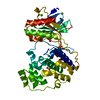
: / Phosphorylase Kinase; domain 1 / Phosphorylase Kinase; domain 1 / Serine/threonine-protein kinase, active site / Serine/Threonine protein kinases active-site signature. / Protein kinase domain / Serine/Threonine protein kinases, catalytic domain / Protein kinase, ATP binding site / Protein kinases ATP-binding region signature. / Protein kinase domain profile. ...: / Phosphorylase Kinase; domain 1 / Phosphorylase Kinase; domain 1 / Serine/threonine-protein kinase, active site / Serine/Threonine protein kinases active-site signature. / Protein kinase domain / Serine/Threonine protein kinases, catalytic domain / Protein kinase, ATP binding site / Protein kinases ATP-binding region signature. / Protein kinase domain profile. / Protein kinase domain / Protein kinase-like domain superfamily / 2-Layer Sandwich / Alpha Beta Similarity search - Domain/homology
Resolution: 2.32→63.39 Å / Cor.coef. Fo:Fc: 0.937 / Cor.coef. Fo:Fc free: 0.925 / SU B: 11.98 / SU ML: 0.147 / Cross valid method: THROUGHOUT / ESU R: 0.267 / ESU R Free: 0.205 / Stereochemistry target values: MAXIMUM LIKELIHOOD / Details: HYDROGENS HAVE BEEN ADDED IN THE RIDING POSITIONS
Rfactor
Num. reflection
% reflection
Selection details
Rfree
0.23797
1195
5.1 %
RANDOM
Rwork
0.21211
-
-
-
obs
0.21345
22080
99.53 %
-
Solvent computation
Ion probe radii: 0.8 Å / Shrinkage radii: 0.8 Å / VDW probe radii: 1.2 Å / Solvent model: MASK
 Movie
Movie Controller


 Open data
 Basic information
 Components
 Keywords
 Function and homology information
 Homo sapiens (human)
 X-RAY DIFFRACTION /
  Authors
 Citation
 Structure visualization
 Downloads & links
 Other downloads


 PDBj
PDBj Assembly


 Mass: 62.068 Da / Num. of mol.: 4 / Source method: obtained synthetically / Formula: C2H6O2
 Mass: 482.617 Da / Num. of mol.: 1 / Source method: obtained synthetically / Formula: C30H34N4O2
 Mass: 18.015 Da / Num. of mol.: 33 / Source method: isolated from a natural source / Formula: H2O
 Sample preparation
 / Beamline: BL13B1 / Wavelength: 1 Å
 Processing