 Movie
Movie Controller

[English] 日本語
 Yorodumi
- PDB-1di9: THE STRUCTURE OF P38 MITOGEN-ACTIVATED PROTEIN KINASE IN COMPLEX ... -
+ Open data

- Basic information
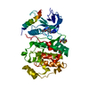
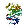
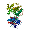
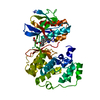
| Entry | Database: PDB / ID: 1di9 | ||||||
|---|---|---|---|---|---|---|---|
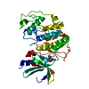
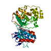
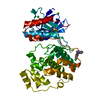
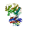
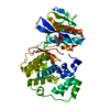
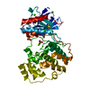
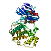
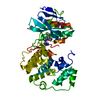
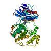
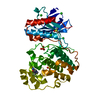
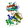
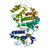
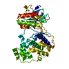
| Title | THE STRUCTURE OF P38 MITOGEN-ACTIVATED PROTEIN KINASE IN COMPLEX WITH 4-[3-METHYLSULFANYLANILINO]-6,7-DIMETHOXYQUINAZOLINE | ||||||
 Components Components | P38 KINASE | ||||||
 Keywords Keywords | TRANSFERASE / SERINE/THREONINE PROTEIN KINASE / MAP KINASE / P38 | ||||||
| Function / homology |  Function and homology information Function and homology informationpositive regulation of cyclase activity / : / Activation of PPARGC1A (PGC-1alpha) by phosphorylation / regulation of synaptic membrane adhesion / stress-induced premature senescence / positive regulation of myoblast fusion / stress-activated protein kinase signaling cascade / CD163 mediating an anti-inflammatory response / 3'-UTR-mediated mRNA stabilization / cell surface receptor protein serine/threonine kinase signaling pathway ...positive regulation of cyclase activity / : / Activation of PPARGC1A (PGC-1alpha) by phosphorylation / regulation of synaptic membrane adhesion / stress-induced premature senescence / positive regulation of myoblast fusion / stress-activated protein kinase signaling cascade / CD163 mediating an anti-inflammatory response / 3'-UTR-mediated mRNA stabilization / cell surface receptor protein serine/threonine kinase signaling pathway / cartilage condensation / positive regulation of myotube differentiation / response to dietary excess / KSRP (KHSRP) binds and destabilizes mRNA / positive regulation of muscle cell differentiation / cellular response to UV-B / mitogen-activated protein kinase p38 binding / Myogenesis / response to muramyl dipeptide / Platelet sensitization by LDL / NFAT protein binding / cellular response to lipoteichoic acid / regulation of cytokine production involved in inflammatory response / Activation of the AP-1 family of transcription factors / p38MAPK cascade / fatty acid oxidation / ERK/MAPK targets / MAP kinase kinase activity / Regulation of MITF-M-dependent genes involved in pigmentation / chondrocyte differentiation / cellular response to vascular endothelial growth factor stimulus / skeletal muscle tissue development / regulation of ossification / MAP kinase activity / mitogen-activated protein kinase / vascular endothelial growth factor receptor signaling pathway / stress-activated MAPK cascade / RHO GTPases Activate NADPH Oxidases / positive regulation of myoblast differentiation / negative regulation of hippo signaling / positive regulation of cardiac muscle cell proliferation / response to muscle stretch / positive regulation of interleukin-12 production / osteoclast differentiation / positive regulation of brown fat cell differentiation / striated muscle cell differentiation / signal transduction in response to DNA damage / lipopolysaccharide-mediated signaling pathway / p38MAPK events / positive regulation of erythrocyte differentiation / placenta development / DNA damage checkpoint signaling / stem cell differentiation / positive regulation of D-glucose import across plasma membrane / tumor necrosis factor-mediated signaling pathway / cellular response to ionizing radiation / activated TAK1 mediates p38 MAPK activation / negative regulation of inflammatory response to antigenic stimulus / negative regulation of canonical Wnt signaling pathway / cellular response to virus / platelet activation / bone development / cell morphogenesis / response to insulin / glucose metabolic process / NOD1/2 Signaling Pathway / positive regulation of protein import into nucleus / VEGFA-VEGFR2 Pathway / cellular senescence / positive regulation of reactive oxygen species metabolic process / chemotaxis / osteoblast differentiation / spindle pole / MAPK cascade / transcription by RNA polymerase II / ADP signalling through P2Y purinoceptor 1 / cellular response to lipopolysaccharide / angiogenesis / secretory granule lumen / protein phosphatase binding / Oxidative Stress Induced Senescence / Regulation of TP53 Activity through Phosphorylation / ficolin-1-rich granule lumen / cell surface receptor signaling pathway / nuclear speck / intracellular signal transduction / protein serine kinase activity / protein serine/threonine kinase activity / positive regulation of gene expression / apoptotic process / regulation of transcription by RNA polymerase II / Neutrophil degranulation / glutamatergic synapse / enzyme binding / signal transduction / positive regulation of transcription by RNA polymerase II / mitochondrion / extracellular region / nucleoplasm / ATP binding Similarity search - Function | ||||||
| Biological species |  Homo sapiens (human) Homo sapiens (human) | ||||||
| Method |  X-RAY DIFFRACTION / Resolution: 2.6 Å X-RAY DIFFRACTION / Resolution: 2.6 Å | ||||||
 Authors Authors | Shewchuk, L. / Hassell, A. / Kuyper, L.F. | ||||||
 Citation Citation | Journal: J.Med.Chem. / Year: 2000 Title: Binding mode of the 4-anilinoquinazoline class of protein kinase inhibitor: X-ray crystallographic studies of 4-anilinoquinazolines bound to cyclin-dependent kinase 2 and p38 kinase. Authors: Shewchuk, L. / Hassell, A. / Wisely, B. / Rocque, W. / Holmes, W. / Veal, J. / Kuyper, L.F. | ||||||
| History |
|
- Structure visualization
| Structure viewer | Molecule: MolmilJmol/JSmol |
|---|
- Downloads & links
-Download
| PDBx/mmCIF format | 1di9.cif.gz | 80.5 KB | Display | PDBx/mmCIF format |
|---|---|---|---|---|
| PDB format | pdb1di9.ent.gz | 61.5 KB | Display | PDB format |
| PDBx/mmJSON format | 1di9.json.gz | Tree view | PDBx/mmJSON format | |
| Others |  Other downloads Other downloads |
-Validation report
| Arichive directory | https://data.pdbj.org/pub/pdb/validation_reports/di/1di9ftp://data.pdbj.org/pub/pdb/validation_reports/di/1di9 | HTTPS FTP |
|---|
-Related structure data


-Links
 PDBj
PDBj


- Assembly
| Deposited unit | 
| ||||||||
|---|---|---|---|---|---|---|---|---|---|
| 1 |
| ||||||||
| Unit cell |
|
-Components
| #1: Protein | Mass: 41343.195 Da / Num. of mol.: 1 Source method: isolated from a genetically manipulated source Source: (gene. exp.) Homo sapiens (human) / Plasmid: PRSETA / Species (production host): Escherichia coli / Production host:  References: UniProt: Q16539, Transferases; Transferring phosphorus-containing groups; Phosphotransferases with an alcohol group as acceptor |
|---|---|
| #2: Chemical | ChemComp-MSQ /   Mass: 327.401 Da / Num. of mol.: 1 / Source method: obtained synthetically / Formula: C17H17N3O2S Mass: 327.401 Da / Num. of mol.: 1 / Source method: obtained synthetically / Formula: C17H17N3O2S |
| #3: Water | ChemComp-HOH /  Mass: 18.015 Da / Num. of mol.: 61 / Source method: isolated from a natural source / Formula: H2O Mass: 18.015 Da / Num. of mol.: 61 / Source method: isolated from a natural source / Formula: H2O |
-Experimental details
-Experiment
| Experiment | Method: X-RAY DIFFRACTION / Number of used crystals: 1 |
|---|
- Sample preparation
| Crystal | Density Matthews: 2.94 Å3/Da / Density % sol: 58.21 % | ||||||||||||||||||||||||||||||||||||||||||||||||||||||||
|---|---|---|---|---|---|---|---|---|---|---|---|---|---|---|---|---|---|---|---|---|---|---|---|---|---|---|---|---|---|---|---|---|---|---|---|---|---|---|---|---|---|---|---|---|---|---|---|---|---|---|---|---|---|---|---|---|---|
| Crystal grow | Temperature: 293 K / Method: vapor diffusion, hanging drop / pH: 7.5 Details: 100MM HEPES, 250MM (NH4)2SO4, 30% PEG6000, pH 7.5, VAPOR DIFFUSION, HANGING DROP, temperature 293K | ||||||||||||||||||||||||||||||||||||||||||||||||||||||||
| Crystal grow | *PLUS Temperature: 20 ℃ / Method: vapor diffusion | ||||||||||||||||||||||||||||||||||||||||||||||||||||||||
| Components of the solutions | *PLUS
|
-Data collection
| Diffraction | Mean temperature: 100 K |
|---|---|
| Diffraction source | Source: ROTATING ANODE / Type: RIGAKU / Wavelength: 1.54 |
| Detector | Type: RIGAKU RAXIS IIC / Detector: IMAGE PLATE / Date: Mar 25, 1998 |
| Radiation | Protocol: SINGLE WAVELENGTH / Monochromatic (M) / Laue (L): M / Scattering type: x-ray |
| Radiation wavelength | Wavelength: 1.54 Å / Relative weight: 1 |
| Reflection | Resolution: 2.6→40 Å / Num. all: 16793 / Num. obs: 16793 / % possible obs: 90.3 % / Observed criterion σ(F): 1 / Observed criterion σ(I): -3 / Redundancy: 5.2 % / Biso Wilson estimate: 18.2 Å2 / Rmerge(I) obs: 0.062 / Net I/σ(I): 30.8 |
| Reflection shell | Resolution: 2.6→2.76 Å / Redundancy: 5 % / Rmerge(I) obs: 0.284 / % possible all: 82.2 |
| Reflection | *PLUS Lowest resolution: 10 Å / Num. obs: 14136 / % possible obs: 90 % / Num. measured all: 88853 / Rmerge(I) obs: 0.065 |
- Processing
| Software |
| ||||||||||||||||||||||||||||||||||||||||||||||||||||||||||||
|---|---|---|---|---|---|---|---|---|---|---|---|---|---|---|---|---|---|---|---|---|---|---|---|---|---|---|---|---|---|---|---|---|---|---|---|---|---|---|---|---|---|---|---|---|---|---|---|---|---|---|---|---|---|---|---|---|---|---|---|---|---|
| Refinement | Resolution: 2.6→40 Å / σ(F): 0 / σ(I): 0 / Stereochemistry target values: ENGH & HUBER
| ||||||||||||||||||||||||||||||||||||||||||||||||||||||||||||
| Refinement step | Cycle: LAST / Resolution: 2.6→40 Å
| ||||||||||||||||||||||||||||||||||||||||||||||||||||||||||||
| Refine LS restraints |
| ||||||||||||||||||||||||||||||||||||||||||||||||||||||||||||
| Software | *PLUS Name: CNS / Classification: refinement | ||||||||||||||||||||||||||||||||||||||||||||||||||||||||||||
| Refinement | *PLUS Highest resolution: 2.6 Å / Lowest resolution: 10 Å / σ(F): 0 / Rfactor obs: 0.206 / Rfactor Rfree: 0.251 | ||||||||||||||||||||||||||||||||||||||||||||||||||||||||||||
| Solvent computation | *PLUS | ||||||||||||||||||||||||||||||||||||||||||||||||||||||||||||
| Displacement parameters | *PLUS |
