 Movie
Movie Controller


+ Open data

- Basic information
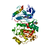
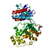
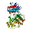
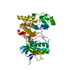
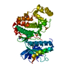
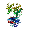
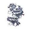
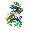
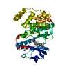
| Entry | Database: PDB / ID: 6qah | ||||||
|---|---|---|---|---|---|---|---|
| Title | ERK2 mini-fragment binding | ||||||
 Components Components | Mitogen-activated protein kinase 1 | ||||||
 Keywords Keywords | SIGNALING PROTEIN / Ser/Thr kinase / signal transduction / ATP binding | ||||||
| Function / homology |  Function and homology information Function and homology informationphospho-PLA2 pathway / Signaling by MAPK mutants / RAF-independent MAPK1/3 activation / Suppression of apoptosis / Gastrin-CREB signalling pathway via PKC and MAPK / Signaling by Activin / cytosine metabolic process / cardiac neural crest cell development involved in heart development / interleukin-34-mediated signaling pathway / caveolin-mediated endocytosis ...phospho-PLA2 pathway / Signaling by MAPK mutants / RAF-independent MAPK1/3 activation / Suppression of apoptosis / Gastrin-CREB signalling pathway via PKC and MAPK / Signaling by Activin / cytosine metabolic process / cardiac neural crest cell development involved in heart development / interleukin-34-mediated signaling pathway / caveolin-mediated endocytosis / response to epidermal growth factor / Signaling by NODAL / Signaling by MAP2K mutants / RSK activation / ERKs are inactivated / Regulation of the apoptosome activity / Golgi Cisternae Pericentriolar Stack Reorganization / positive regulation of macrophage proliferation / outer ear morphogenesis / : / regulation of Golgi inheritance / positive regulation of peptidyl-threonine phosphorylation / Signaling by LTK in cancer / labyrinthine layer blood vessel development / mammary gland epithelial cell proliferation / ERBB signaling pathway / trachea formation / regulation of early endosome to late endosome transport / Negative feedback regulation of MAPK pathway / regulation of stress-activated MAPK cascade / IFNG signaling activates MAPKs / positive regulation of neuroinflammatory response / Frs2-mediated activation / Activation of the AP-1 family of transcription factors / ERBB2-ERBB3 signaling pathway / response to exogenous dsRNA / RUNX2 regulates osteoblast differentiation / ERK/MAPK targets / regulation of cytoskeleton organization / MAPK1 (ERK2) activation / positive regulation of macrophage chemotaxis / face development / Bergmann glial cell differentiation / Recycling pathway of L1 / thyroid gland development / pseudopodium / lung morphogenesis / positive regulation of telomere maintenance / MAP kinase activity / regulation of ossification / Advanced glycosylation endproduct receptor signaling / mitogen-activated protein kinase / Regulation of HSF1-mediated heat shock response / Estrogen-dependent nuclear events downstream of ESR-membrane signaling / negative regulation of cell differentiation / RHO GTPases Activate NADPH Oxidases / Signal attenuation / RHO GTPases Activate WASPs and WAVEs / Growth hormone receptor signaling / Schwann cell development / stress-activated MAPK cascade / phosphatase binding / Estrogen-stimulated signaling through PRKCZ / ERK1 and ERK2 cascade / NPAS4 regulates expression of target genes / phosphotyrosine residue binding / Nuclear events stimulated by ALK signaling in cancer / myelination / RNA polymerase II CTD heptapeptide repeat kinase activity / Transcriptional and post-translational regulation of MITF-M expression and activity / ESR-mediated signaling / NCAM signaling for neurite out-growth / insulin-like growth factor receptor signaling pathway / lipopolysaccharide-mediated signaling pathway / thymus development / ciliary tip / cellular response to amino acid starvation / Regulation of PTEN gene transcription / Signal transduction by L1 / B cell receptor signaling pathway / chemokine-mediated signaling pathway / response to nicotine / FCGR3A-mediated phagocytosis / FCERI mediated MAPK activation / Negative regulation of FGFR3 signaling / Negative regulation of FGFR2 signaling / Negative regulation of FGFR4 signaling / Downregulation of SMAD2/3:SMAD4 transcriptional activity / Negative regulation of FGFR1 signaling / SMAD2/SMAD3:SMAD4 heterotrimer regulates transcription / Signaling by high-kinase activity BRAF mutants / Spry regulation of FGF signaling / MAP2K and MAPK activation / positive regulation of cholesterol biosynthetic process / Regulation of actin dynamics for phagocytic cup formation / Negative Regulation of CDH1 Gene Transcription / Oncogene Induced Senescence / caveola / cellular response to tumor necrosis factor / epidermal growth factor receptor signaling pathway Similarity search - Function | ||||||
| Biological species |  Homo sapiens (human) Homo sapiens (human) | ||||||
| Method |  X-RAY DIFFRACTION / FOURIER SYNTHESIS / Resolution: 1.58 Å X-RAY DIFFRACTION / FOURIER SYNTHESIS / Resolution: 1.58 Å | ||||||
 Authors Authors | O'Reilly, M. / Cleasby, A. / Davies, T.G. / Hall, R. / Ludlow, F. / Murray, C.W. / Tisi, D. / Jhoti, H. | ||||||
 Citation Citation | Journal: Drug Discov Today / Year: 2019 Title: Crystallographic screening using ultra-low-molecular-weight ligands to guide drug design. Authors: O'Reilly, M. / Cleasby, A. / Davies, T.G. / Hall, R.J. / Ludlow, R.F. / Murray, C.W. / Tisi, D. / Jhoti, H. | ||||||
| History |
|
- Structure visualization
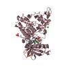
| Structure viewer | Molecule: MolmilJmol/JSmol |
|---|
- Downloads & links
-Download
| PDBx/mmCIF format | 6qah.cif.gz | 159.7 KB | Display | PDBx/mmCIF format |
|---|---|---|---|---|
| PDB format | pdb6qah.ent.gz | 123.4 KB | Display | PDB format |
| PDBx/mmJSON format | 6qah.json.gz | Tree view | PDBx/mmJSON format | |
| Others |  Other downloads Other downloads |
-Validation report
| Arichive directory | https://data.pdbj.org/pub/pdb/validation_reports/qa/6qahftp://data.pdbj.org/pub/pdb/validation_reports/qa/6qah | HTTPS FTP |
|---|
-Related structure data
| Related structure data |  6q7kC 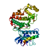 6q7sC  6q7tC  6qa1C  6qa3C  6qa4C  6qagC  6qalC  6qaqC  6qawC C: citing same article ( |
|---|---|
| Similar structure data |


-Links
 PDBj
PDBj


- Assembly
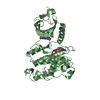
| Deposited unit | 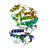
| ||||||||
|---|---|---|---|---|---|---|---|---|---|
| 1 |
| ||||||||
| Unit cell |
|
-Components
| #1: Protein | Mass: 42551.922 Da / Num. of mol.: 1 Source method: isolated from a genetically manipulated source Source: (gene. exp.) Homo sapiens (human) / Gene: MAPK1, ERK2, PRKM1, PRKM2 / Production host:  References: UniProt: P28482, mitogen-activated protein kinase | ||||
|---|---|---|---|---|---|
| #2: Chemical | ChemComp-SO4 /   Mass: 96.063 Da / Num. of mol.: 1 / Source method: obtained synthetically / Formula: SO4 Mass: 96.063 Da / Num. of mol.: 1 / Source method: obtained synthetically / Formula: SO4 | ||||
| #3: Chemical | 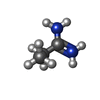  Mass: 73.117 Da / Num. of mol.: 2 / Source method: obtained synthetically / Formula: C3H9N2 Mass: 73.117 Da / Num. of mol.: 2 / Source method: obtained synthetically / Formula: C3H9N2#4: Water | ChemComp-HOH / |  Mass: 18.015 Da / Num. of mol.: 244 / Source method: isolated from a natural source / Formula: H2O Mass: 18.015 Da / Num. of mol.: 244 / Source method: isolated from a natural source / Formula: H2OHas protein modification | Y | |
-Experimental details
-Experiment
| Experiment | Method: X-RAY DIFFRACTION / Number of used crystals: 1 |
|---|
- Sample preparation
| Crystal | Density Matthews: 2.29 Å3/Da / Density % sol: 46.38 % |
|---|---|
| Crystal grow | Temperature: 293 K / Method: vapor diffusion, sitting drop / pH: 7.2 Details: 0.2M (NH4)2SO4 34% MPEG 2000 0.02M Mercaptoethanol 0.1M pH=7.2 HEPES/NaOH |
-Data collection
| Diffraction | Mean temperature: 100 K / Serial crystal experiment: N |
|---|---|
| Diffraction source | Source: ROTATING ANODE / Type: RIGAKU FR-X / Wavelength: 1.54178 Å |
| Detector | Type: DECTRIS PILATUS 300K / Detector: PIXEL / Date: Apr 7, 2017 |
| Radiation | Protocol: SINGLE WAVELENGTH / Monochromatic (M) / Laue (L): M / Scattering type: x-ray |
| Radiation wavelength | Wavelength: 1.54178 Å / Relative weight: 1 |
| Reflection | Resolution: 1.58→48.74 Å / Num. obs: 37802 / % possible obs: 72.4 % / Redundancy: 3.1 % / Biso Wilson estimate: 21.81 Å2 / Rrim(I) all: 0.037 / Net I/σ(I): 15.5 |
| Reflection shell | Resolution: 1.58→1.61 Å / Num. unique obs: 118 / Rrim(I) all: 1.261 / % possible all: 6.3 |
- Processing
| Software |
| ||||||||||||||||||||||||||||||||||||||||||||||||||||||||||||||||||||||||||||||||||||||||||||||||||||||||||||||||||
|---|---|---|---|---|---|---|---|---|---|---|---|---|---|---|---|---|---|---|---|---|---|---|---|---|---|---|---|---|---|---|---|---|---|---|---|---|---|---|---|---|---|---|---|---|---|---|---|---|---|---|---|---|---|---|---|---|---|---|---|---|---|---|---|---|---|---|---|---|---|---|---|---|---|---|---|---|---|---|---|---|---|---|---|---|---|---|---|---|---|---|---|---|---|---|---|---|---|---|---|---|---|---|---|---|---|---|---|---|---|---|---|---|---|---|---|
| Refinement | Method to determine structure: FOURIER SYNTHESIS / Resolution: 1.58→34.89 Å / Cor.coef. Fo:Fc: 0.949 / Cor.coef. Fo:Fc free: 0.933 / SU R Cruickshank DPI: 0.149 / Cross valid method: THROUGHOUT / σ(F): 0 / SU R Blow DPI: 0.112 / SU Rfree Blow DPI: 0.107 / SU Rfree Cruickshank DPI: 0.106
| ||||||||||||||||||||||||||||||||||||||||||||||||||||||||||||||||||||||||||||||||||||||||||||||||||||||||||||||||||
| Displacement parameters | Biso mean: 29.596 Å2
| ||||||||||||||||||||||||||||||||||||||||||||||||||||||||||||||||||||||||||||||||||||||||||||||||||||||||||||||||||
| Refine analyze | Luzzati coordinate error obs: 0.21 Å | ||||||||||||||||||||||||||||||||||||||||||||||||||||||||||||||||||||||||||||||||||||||||||||||||||||||||||||||||||
| Refinement step | Cycle: 1 / Resolution: 1.58→34.89 Å
| ||||||||||||||||||||||||||||||||||||||||||||||||||||||||||||||||||||||||||||||||||||||||||||||||||||||||||||||||||
| Refine LS restraints |
| ||||||||||||||||||||||||||||||||||||||||||||||||||||||||||||||||||||||||||||||||||||||||||||||||||||||||||||||||||
| LS refinement shell | Resolution: 1.58→1.68 Å / Total num. of bins used: 50
| ||||||||||||||||||||||||||||||||||||||||||||||||||||||||||||||||||||||||||||||||||||||||||||||||||||||||||||||||||
| Refinement TLS params. | Method: refined / Origin x: -1.3833 Å / Origin y: 3.3211 Å / Origin z: 37.7164 Å
| ||||||||||||||||||||||||||||||||||||||||||||||||||||||||||||||||||||||||||||||||||||||||||||||||||||||||||||||||||
| Refinement TLS group | Selection details: { A|11 - A|365 } |
