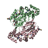
G-protein-coupled receptor kinase / G protein-coupled receptor kinase activity / G alpha (s) signalling events / regulation of signal transduction / Wnt signaling pathway / protein autophosphorylation / protein kinase activity / protein serine/threonine kinase activity / apoptotic process / lipid binding ...G-protein-coupled receptor kinase / G protein-coupled receptor kinase activity / G alpha (s) signalling events / regulation of signal transduction / Wnt signaling pathway / protein autophosphorylation / protein kinase activity / protein serine/threonine kinase activity / apoptotic process / lipid binding / negative regulation of apoptotic process / ATP binding / nucleus / plasma membrane / cytoplasm Similarity search - Function
GPCR kinase / Regulator of G-protein Signalling 4, domain 2 / Regulator of G-protein Signalling 4; domain 2 / Regulator of G protein signaling domain / RGS domain / RGS domain profile. / Regulator of G protein signalling domain / RGS, subdomain 2 / RGS domain superfamily / Extension to Ser/Thr-type protein kinases ...GPCR kinase / Regulator of G-protein Signalling 4, domain 2 / Regulator of G-protein Signalling 4; domain 2 / Regulator of G protein signaling domain / RGS domain / RGS domain profile. / Regulator of G protein signalling domain / RGS, subdomain 2 / RGS domain superfamily / Extension to Ser/Thr-type protein kinases / AGC-kinase, C-terminal / AGC-kinase C-terminal domain profile. / Phosphorylase Kinase; domain 1 / Phosphorylase Kinase; domain 1 / Transferase(Phosphotransferase) domain 1 / Transferase(Phosphotransferase); domain 1 / Serine/threonine-protein kinase, active site / Serine/Threonine protein kinases active-site signature. / Protein kinase domain / Serine/Threonine protein kinases, catalytic domain / Protein kinase, ATP binding site / Protein kinases ATP-binding region signature. / Protein kinase domain profile. / Protein kinase domain / Protein kinase-like domain superfamily / 2-Layer Sandwich / Orthogonal Bundle / Mainly Alpha / Alpha Beta Similarity search - Domain/homology
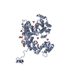
Resolution: 2.42→24.47 Å / Cor.coef. Fo:Fc: 0.962 / Cor.coef. Fo:Fc free: 0.942 / SU B: 20.69 / SU ML: 0.212 / Cross valid method: THROUGHOUT / ESU R: 0.39 / ESU R Free: 0.256 / Stereochemistry target values: MAXIMUM LIKELIHOOD / Details: HYDROGENS HAVE BEEN ADDED IN THE RIDING POSITIONS
Rfactor
Num. reflection
% reflection
Selection details
Rfree
0.24297
1235
5.1 %
RANDOM
Rwork
0.19379
-
-
-
obs
0.19635
22906
99.56 %
-
Solvent computation
Ion probe radii: 0.8 Å / Shrinkage radii: 0.8 Å / VDW probe radii: 1.2 Å / Solvent model: MASK
 Movie
Movie Controller

 Yorodumi
 Open data
 Basic information
 Components
 Keywords
 Function and homology information
 X-RAY DIFFRACTION /
  Authors
 United States, 3items
  Citation
 Structure visualization
 Downloads & links
 Other downloads
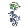


 PDBj
PDBj


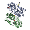
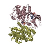
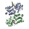
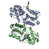
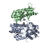
 Assembly
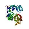
 Trichoplusia ni (cabbage looper)
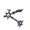
 Mass: 499.496 Da / Num. of mol.: 1 / Source method: obtained synthetically / Formula: C26H22FN7O3
 Mass: 96.063 Da / Num. of mol.: 1 / Source method: obtained synthetically / Formula: SO4
 Mass: 18.015 Da / Num. of mol.: 37 / Source method: isolated from a natural source / Formula: H2O
 Sample preparation
 Processing