- PDB-4uzd: SAR156497 an exquisitely selective inhibitor of Aurora kinases -
+
Open data
ID or keywords:
Loading...
-
Basic information
Entry
Database: PDB / ID: 4uzd
Title
SAR156497 an exquisitely selective inhibitor of Aurora kinases
Components
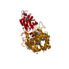
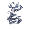
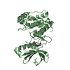
AURORA KINASE A
Keywords
TRANSFERASE
Function / homology
Function and homology information
Interaction between PHLDA1 and AURKA / regulation of centrosome cycle / axon hillock / spindle assembly involved in female meiosis I / cilium disassembly / spindle pole centrosome / mitotic centrosome separation / histone H3S10 kinase activity / chromosome passenger complex / positive regulation of oocyte maturation ...Interaction between PHLDA1 and AURKA / regulation of centrosome cycle / axon hillock / spindle assembly involved in female meiosis I / cilium disassembly / spindle pole centrosome / mitotic centrosome separation / histone H3S10 kinase activity / chromosome passenger complex / positive regulation of oocyte maturation / pronucleus / germinal vesicle / protein localization to centrosome / meiotic spindle / anterior/posterior axis specification / neuron projection extension / centrosome localization / spindle organization / positive regulation of mitochondrial fission / mitotic spindle pole / spindle midzone / SUMOylation of DNA replication proteins / negative regulation of protein binding / regulation of G2/M transition of mitotic cell cycle / positive regulation of mitotic cell cycle / positive regulation of mitotic nuclear division / protein serine/threonine/tyrosine kinase activity / centriole / TP53 Regulates Transcription of Genes Involved in G2 Cell Cycle Arrest / liver regeneration / AURKA Activation by TPX2 / molecular function activator activity / regulation of signal transduction by p53 class mediator / mitotic spindle organization / peptidyl-serine phosphorylation / regulation of cytokinesis / regulation of protein stability / response to wounding / APC/C:Cdh1 mediated degradation of Cdc20 and other APC/C:Cdh1 targeted proteins in late mitosis/early G1 / FBXL7 down-regulates AURKA during mitotic entry and in early mitosis / G2/M transition of mitotic cell cycle / kinetochore / spindle / spindle pole / mitotic spindle / Regulation of PLK1 Activity at G2/M Transition / protein autophosphorylation / mitotic cell cycle / positive regulation of proteasomal ubiquitin-dependent protein catabolic process / microtubule cytoskeleton / midbody / Regulation of TP53 Activity through Phosphorylation / basolateral plasma membrane / microtubule / proteasome-mediated ubiquitin-dependent protein catabolic process / protein phosphorylation / protein kinase activity / non-specific serine/threonine protein kinase / ciliary basal body / postsynaptic density / protein heterodimerization activity / negative regulation of gene expression / protein serine kinase activity / cell division / protein serine/threonine kinase activity / apoptotic process / centrosome / ubiquitin protein ligase binding / protein kinase binding / negative regulation of apoptotic process / perinuclear region of cytoplasm / glutamatergic synapse / nucleoplasm / ATP binding / nucleus / cytosol Similarity search - Function
Aurora kinase A / Aurora kinase / Phosphorylase Kinase; domain 1 / Phosphorylase Kinase; domain 1 / Transferase(Phosphotransferase) domain 1 / Transferase(Phosphotransferase); domain 1 / Serine/threonine-protein kinase, active site / Serine/Threonine protein kinases active-site signature. / Protein kinase domain / Serine/Threonine protein kinases, catalytic domain ...Aurora kinase A / Aurora kinase / Phosphorylase Kinase; domain 1 / Phosphorylase Kinase; domain 1 / Transferase(Phosphotransferase) domain 1 / Transferase(Phosphotransferase); domain 1 / Serine/threonine-protein kinase, active site / Serine/Threonine protein kinases active-site signature. / Protein kinase domain / Serine/Threonine protein kinases, catalytic domain / Protein kinase, ATP binding site / Protein kinases ATP-binding region signature. / Protein kinase domain profile. / Protein kinase domain / Protein kinase-like domain superfamily / 2-Layer Sandwich / Orthogonal Bundle / Mainly Alpha / Alpha Beta Similarity search - Domain/homology
Protocol: SINGLE WAVELENGTH / Monochromatic (M) / Laue (L): M / Scattering type: x-ray
Radiation wavelength
Wavelength: 0.9762 Å / Relative weight: 1
Reflection
Resolution: 3.2→78.3 Å / Num. obs: 16476 / % possible obs: 97.9 % / Observed criterion σ(I): 2 / Redundancy: 5.8 % / Biso Wilson estimate: 117.47 Å2 / Rmerge(I) obs: 0.08 / Net I/σ(I): 19.9
Reflection shell
Resolution: 3.2→3.37 Å / Redundancy: 5.9 % / Rmerge(I) obs: 0.41 / Mean I/σ(I) obs: 3.1 / % possible all: 99.3
-
Processing
Software
Name
Version
Classification
BUSTER
2.11.5
refinement
MOSFLM
datareduction
SCALA
datascaling
PHASER
phasing
Refinement
Method to determine structure: MOLECULAR REPLACEMENT Starting model: NONE Resolution: 3.2→78.33 Å / Cor.coef. Fo:Fc: 0.9142 / Cor.coef. Fo:Fc free: 0.8679 / Cross valid method: THROUGHOUT / σ(F): 0 / SU Rfree Blow DPI: 0.423 Details: IDEAL-DIST CONTACT TERM CONTACT SETUP. ALL ATOMS HAVE CCP4 ATOM TYPE FROM LIBRARY
Rfactor
Num. reflection
% reflection
Selection details
Rfree
0.262
1270
7.73 %
RANDOM
Rwork
0.215
-
-
-
obs
-
16434
97.61 %
-
Displacement parameters
Biso mean: 94.63 Å2
Baniso -1
Baniso -2
Baniso -3
1-
-11.2179 Å2
0 Å2
0 Å2
2-
-
-11.2179 Å2
0 Å2
3-
-
-
22.4359 Å2
Refine analyze
Luzzati coordinate error obs: 0.707 Å
Refinement step
Cycle: LAST / Resolution: 3.2→78.33 Å
Protein
Nucleic acid
Ligand
Solvent
Total
Num. atoms
4217
0
70
0
4287
Refine LS restraints
Refine-ID
Type
Dev ideal
Number
Restraint function
Weight
X-RAY DIFFRACTION
t_bond_d
0.01
4397
HARMONIC
2
X-RAY DIFFRACTION
t_angle_deg
1.05
5943
HARMONIC
2
X-RAY DIFFRACTION
t_dihedral_angle_d
1577
SINUSOIDAL
2
X-RAY DIFFRACTION
t_incorr_chiral_ct
X-RAY DIFFRACTION
t_pseud_angle
X-RAY DIFFRACTION
t_trig_c_planes
98
HARMONIC
2
X-RAY DIFFRACTION
t_gen_planes
700
HARMONIC
5
X-RAY DIFFRACTION
t_it
4397
HARMONIC
20
X-RAY DIFFRACTION
t_nbd
X-RAY DIFFRACTION
t_omega_torsion
2.59
X-RAY DIFFRACTION
t_other_torsion
18.64
X-RAY DIFFRACTION
t_improper_torsion
X-RAY DIFFRACTION
t_chiral_improper_torsion
535
SEMIHARMONIC
5
X-RAY DIFFRACTION
t_sum_occupancies
X-RAY DIFFRACTION
t_utility_distance
X-RAY DIFFRACTION
t_utility_angle
X-RAY DIFFRACTION
t_utility_torsion
X-RAY DIFFRACTION
t_ideal_dist_contact
5088
SEMIHARMONIC
4
LS refinement shell
Resolution: 3.2→3.42 Å / Total num. of bins used: 8
Rfactor
Num. reflection
% reflection
Rfree
0.2827
255
8.63 %
Rwork
0.2577
2701
-
all
0.2598
2956
-
obs
-
-
97.61 %
+
About Yorodumi
-
News
-
Feb 9, 2022. New format data for meta-information of EMDB entries
New format data for meta-information of EMDB entries
Version 3 of the EMDB header file is now the official format.
The previous official version 1.9 will be removed from the archive.
In the structure databanks used in Yorodumi, some data are registered as the other names, "COVID-19 virus" and "2019-nCoV". Here are the details of the virus and the list of structure data.
Jan 31, 2019. EMDB accession codes are about to change! (news from PDBe EMDB page)
EMDB accession codes are about to change! (news from PDBe EMDB page)
The allocation of 4 digits for EMDB accession codes will soon come to an end. Whilst these codes will remain in use, new EMDB accession codes will include an additional digit and will expand incrementally as the available range of codes is exhausted. The current 4-digit format prefixed with “EMD-” (i.e. EMD-XXXX) will advance to a 5-digit format (i.e. EMD-XXXXX), and so on. It is currently estimated that the 4-digit codes will be depleted around Spring 2019, at which point the 5-digit format will come into force.
The EM Navigator/Yorodumi systems omit the EMD- prefix.
Related info.:Q: What is EMD? / ID/Accession-code notation in Yorodumi/EM Navigator
Yorodumi is a browser for structure data from EMDB, PDB, SASBDB, etc.
This page is also the successor to EM Navigator detail page, and also detail information page/front-end page for Omokage search.
The word "yorodu" (or yorozu) is an old Japanese word meaning "ten thousand". "mi" (miru) is to see.
Related info.:EMDB / PDB / SASBDB / Comparison of 3 databanks / Yorodumi Search / Aug 31, 2016. New EM Navigator & Yorodumi / Yorodumi Papers / Jmol/JSmol / Function and homology information / Changes in new EM Navigator and Yorodumi
 Movie
Movie Controller


 Open data
 Basic information
 Components
 Keywords
 Function and homology information
 HOMO SAPIENS (human)
 X-RAY DIFFRACTION /
  Authors
 Citation
 Structure visualization
 Downloads & links
 Other downloads


 PDBj
PDBj


 Assembly


 Mass: 468.504 Da / Num. of mol.: 2 / Source method: obtained synthetically / Formula: C27H24N4O4
 Sample preparation
 / Beamline: ID29 / Wavelength: 0.9762
  Processing