 Movie
Movie Controller

[English] 日本語
 Yorodumi
- PDB-5xo1: Crystal structure of the isochorismatase domain of VabB from Vibr... -
+ Open data

- Basic information
| Entry | Database: PDB / ID: 5xo1 | |||||||||||||||
|---|---|---|---|---|---|---|---|---|---|---|---|---|---|---|---|---|
| Title | Crystal structure of the isochorismatase domain of VabB from Vibrio anguillarum 775 | |||||||||||||||
 Components Components | Isochorismate lyase | |||||||||||||||
 Keywords Keywords | HYDROLASE / VabB / isochorismatase / siderophore / iron | |||||||||||||||
| Function / homology |  Function and homology information Function and homology information | |||||||||||||||
| Biological species |  Vibrio anguillarum 775 (bacteria) Vibrio anguillarum 775 (bacteria) | |||||||||||||||
| Method |  X-RAY DIFFRACTION / SYNCHROTRON / MOLECULAR REPLACEMENT / Resolution: 2.23 Å X-RAY DIFFRACTION / SYNCHROTRON / MOLECULAR REPLACEMENT / Resolution: 2.23 Å | |||||||||||||||
 Authors Authors | Ma, Q. / Du, J. | |||||||||||||||
| Funding support |  China, 4items China, 4items
| |||||||||||||||
 Citation Citation | Journal: Biochem.Biophys.Res.Commun. / Year: 2017 Title: Crystal structures of the isochorismatase domains from Vibrio anguillarum. Authors: Du, J. / Deng, T. / Ma, Q. | |||||||||||||||
| History |
|
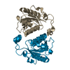
- Structure visualization
| Structure viewer | Molecule: MolmilJmol/JSmol |
|---|
- Downloads & links
-Download
| PDBx/mmCIF format | 5xo1.cif.gz | 494.3 KB | Display | PDBx/mmCIF format |
|---|---|---|---|---|
| PDB format | pdb5xo1.ent.gz | 411.2 KB | Display | PDB format |
| PDBx/mmJSON format | 5xo1.json.gz | Tree view | PDBx/mmJSON format | |
| Others |  Other downloads Other downloads |
-Validation report
| Arichive directory | https://data.pdbj.org/pub/pdb/validation_reports/xo/5xo1ftp://data.pdbj.org/pub/pdb/validation_reports/xo/5xo1 | HTTPS FTP |
|---|
-Related structure data
| Related structure data |  5xo0SC S: Starting model for refinement C: citing same article ( |
|---|---|
| Similar structure data |


-Links
 PDBj
PDBj
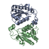
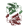
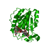
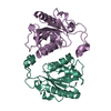
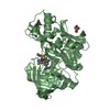
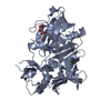
- Assembly
| Deposited unit | 
| ||||||||
|---|---|---|---|---|---|---|---|---|---|
| 1 | 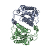
| ||||||||
| 2 | 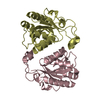
| ||||||||
| 3 | 
| ||||||||
| Unit cell |
|
-Components
| #1: Protein | Mass: 24782.371 Da / Num. of mol.: 6 / Fragment: UNP residues 1-217 Source method: isolated from a genetically manipulated source Source: (gene. exp.) Vibrio anguillarum 775 (bacteria) / Strain: 775 / Gene: vabB / Production host: #2: Water | ChemComp-HOH / |  Mass: 18.015 Da / Num. of mol.: 175 / Source method: isolated from a natural source / Formula: H2O Mass: 18.015 Da / Num. of mol.: 175 / Source method: isolated from a natural source / Formula: H2O |
|---|
-Experimental details
-Experiment
| Experiment | Method: X-RAY DIFFRACTION / Number of used crystals: 1 |
|---|
- Sample preparation
| Crystal | Density Matthews: 2.18 Å3/Da / Density % sol: 43.53 % |
|---|---|
| Crystal grow | Temperature: 293 K / Method: vapor diffusion, sitting drop / Details: 10% PEG 8000, 0.1M imidazole, 0.2M calcium acetate |
-Data collection
| Diffraction | Mean temperature: 100 K |
|---|---|
| Diffraction source | Source: SYNCHROTRON / Site: SSRF / Beamline: BL17U1 / Wavelength: 0.9791 Å |
| Detector | Type: ADSC QUANTUM 315r / Detector: CCD / Date: Nov 15, 2016 / Details: dynamically bendable toroidal mirror |
| Radiation | Monochromator: double crystal monochromator / Protocol: SINGLE WAVELENGTH / Monochromatic (M) / Laue (L): M / Scattering type: x-ray |
| Radiation wavelength | Wavelength: 0.9791 Å / Relative weight: 1 |
| Reflection | Resolution: 2.23→143.85 Å / Num. obs: 61526 / % possible obs: 99.6 % / Redundancy: 14 % / Biso Wilson estimate: 49.4 Å2 / CC1/2: 0.996 / Rmerge(I) obs: 0.12 / Rpim(I) all: 0.033 / Rsym value: 0.12 / Net I/σ(I): 14.5 |
| Reflection shell | Resolution: 2.23→2.238 Å / Redundancy: 11.8 % / Rmerge(I) obs: 1.189 / Mean I/σ(I) obs: 1.9 / Num. unique obs: 588 / CC1/2: 0.627 / Rpim(I) all: 0.357 / Rsym value: 1.189 / % possible all: 90.3 |
- Processing
| Software |
| |||||||||||||||||||||||||||||||||||||||||||||||||||||||||||||||||||||||||||||||||||||||||||||||||||||||||||||||||||||||||||||||||||||||||||||||||||||||||||||||||||||||||||||||
|---|---|---|---|---|---|---|---|---|---|---|---|---|---|---|---|---|---|---|---|---|---|---|---|---|---|---|---|---|---|---|---|---|---|---|---|---|---|---|---|---|---|---|---|---|---|---|---|---|---|---|---|---|---|---|---|---|---|---|---|---|---|---|---|---|---|---|---|---|---|---|---|---|---|---|---|---|---|---|---|---|---|---|---|---|---|---|---|---|---|---|---|---|---|---|---|---|---|---|---|---|---|---|---|---|---|---|---|---|---|---|---|---|---|---|---|---|---|---|---|---|---|---|---|---|---|---|---|---|---|---|---|---|---|---|---|---|---|---|---|---|---|---|---|---|---|---|---|---|---|---|---|---|---|---|---|---|---|---|---|---|---|---|---|---|---|---|---|---|---|---|---|---|---|---|---|---|
| Refinement | Method to determine structure: MOLECULAR REPLACEMENT Starting model: 5XO0 Resolution: 2.23→32.65 Å / Cor.coef. Fo:Fc: 0.938 / Cor.coef. Fo:Fc free: 0.939 / Rfactor Rfree error: 0 / SU R Cruickshank DPI: 0.291 / Cross valid method: THROUGHOUT / σ(F): 0 / SU R Blow DPI: 0.286 / SU Rfree Blow DPI: 0.187 / SU Rfree Cruickshank DPI: 0.19 Details: TLS parameters are refined for each chain. Thin shell test set is used.
| |||||||||||||||||||||||||||||||||||||||||||||||||||||||||||||||||||||||||||||||||||||||||||||||||||||||||||||||||||||||||||||||||||||||||||||||||||||||||||||||||||||||||||||||
| Displacement parameters | Biso mean: 58.4 Å2
| |||||||||||||||||||||||||||||||||||||||||||||||||||||||||||||||||||||||||||||||||||||||||||||||||||||||||||||||||||||||||||||||||||||||||||||||||||||||||||||||||||||||||||||||
| Refine analyze | Luzzati coordinate error obs: 0.31 Å | |||||||||||||||||||||||||||||||||||||||||||||||||||||||||||||||||||||||||||||||||||||||||||||||||||||||||||||||||||||||||||||||||||||||||||||||||||||||||||||||||||||||||||||||
| Refinement step | Cycle: 1 / Resolution: 2.23→32.65 Å
| |||||||||||||||||||||||||||||||||||||||||||||||||||||||||||||||||||||||||||||||||||||||||||||||||||||||||||||||||||||||||||||||||||||||||||||||||||||||||||||||||||||||||||||||
| Refine LS restraints |
| |||||||||||||||||||||||||||||||||||||||||||||||||||||||||||||||||||||||||||||||||||||||||||||||||||||||||||||||||||||||||||||||||||||||||||||||||||||||||||||||||||||||||||||||
| LS refinement shell | Resolution: 2.23→2.29 Å / Total num. of bins used: 20
| |||||||||||||||||||||||||||||||||||||||||||||||||||||||||||||||||||||||||||||||||||||||||||||||||||||||||||||||||||||||||||||||||||||||||||||||||||||||||||||||||||||||||||||||
| Refinement TLS params. | Method: refined / Refine-ID: X-RAY DIFFRACTION
| |||||||||||||||||||||||||||||||||||||||||||||||||||||||||||||||||||||||||||||||||||||||||||||||||||||||||||||||||||||||||||||||||||||||||||||||||||||||||||||||||||||||||||||||
| Refinement TLS group |
|
