 Movie
Movie Controller


+ Open data

- Basic information
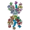
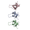
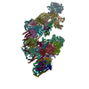
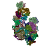
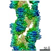
| Entry | Database: PDB / ID: 4uf9 | ||||||
|---|---|---|---|---|---|---|---|
| Title | Electron cryo-microscopy structure of PB1-p62 type T filaments | ||||||
 Components Components | SEQUESTOSOME-1 | ||||||
 Keywords Keywords | SIGNALING PROTEIN / SELECTIVE AUTOPHAGY / AUTOPHAGY RECEPTOR / AUTOPHAGY SCAFFOLD / P62/SQSTM1 / SINGLE-PARTICLE HELICAL RECONSTRUCTION | ||||||
| Function / homology |  Function and homology information Function and homology informationprotein localization to perinuclear region of cytoplasm / brown fat cell proliferation / protein targeting to vacuole involved in autophagy / regulation of Ras protein signal transduction / intracellular membraneless organelle / aggrephagy / negative regulation of toll-like receptor 4 signaling pathway / response to mitochondrial depolarisation / amphisome / regulation of protein complex stability ...protein localization to perinuclear region of cytoplasm / brown fat cell proliferation / protein targeting to vacuole involved in autophagy / regulation of Ras protein signal transduction / intracellular membraneless organelle / aggrephagy / negative regulation of toll-like receptor 4 signaling pathway / response to mitochondrial depolarisation / amphisome / regulation of protein complex stability / autophagy of mitochondrion / endosome organization / pexophagy / membraneless organelle assembly / regulation of mitochondrion organization / phagophore assembly site / ubiquitin-modified protein reader activity / aggresome / Nuclear events mediated by NFE2L2 / regulation of canonical NF-kappaB signal transduction / K63-linked polyubiquitin modification-dependent protein binding / endosomal transport / cellular response to stress / Lewy body / temperature homeostasis / autolysosome / negative regulation of ferroptosis / molecular sequestering activity / energy homeostasis / mitophagy / immune system process / inclusion body / signaling adaptor activity / negative regulation of protein ubiquitination / positive regulation of autophagy / ionotropic glutamate receptor binding / autophagosome / SH2 domain binding / response to ischemia / p75NTR recruits signalling complexes / protein catabolic process / NF-kB is activated and signals survival / Pexophagy / protein kinase C binding / NRIF signals cell death from the nucleus / positive regulation of long-term synaptic potentiation / positive regulation of protein localization to plasma membrane / PINK1-PRKN Mediated Mitophagy / sarcomere / macroautophagy / ubiquitin binding / protein sequestering activity / molecular condensate scaffold activity / P-body / receptor tyrosine kinase binding / PML body / protein import into nucleus / autophagy / Interleukin-1 signaling / intracellular protein localization / Signaling by ALK fusions and activated point mutants / late endosome / KEAP1-NFE2L2 pathway / sperm midpiece / signaling receptor activity / Neddylation / ubiquitin-dependent protein catabolic process / protein-macromolecule adaptor activity / cell differentiation / intracellular signal transduction / positive regulation of apoptotic process / apoptotic process / ubiquitin protein ligase binding / protein kinase binding / protein-containing complex binding / glutamatergic synapse / enzyme binding / endoplasmic reticulum / positive regulation of transcription by RNA polymerase II / mitochondrion / extracellular exosome / zinc ion binding / nucleoplasm / identical protein binding / cytosol / cytoplasm Similarity search - Function | ||||||
| Biological species |  HOMO SAPIENS (human) HOMO SAPIENS (human) | ||||||
| Method | ELECTRON MICROSCOPY / helical reconstruction / cryo EM / Resolution: 10.3 Å | ||||||
 Authors Authors | Ciuffa, R. / Lamark, T. / Tarafder, A. / Guesdon, A. / Rybina, S. / Hagen, W.J.H. / Johansen, T. / Sachse, C. | ||||||
 Citation Citation | Journal: Cell Rep / Year: 2015 Title: The selective autophagy receptor p62 forms a flexible filamentous helical scaffold. Authors: Rodolfo Ciuffa / Trond Lamark / Abul K Tarafder / Audrey Guesdon / Sofia Rybina / Wim J H Hagen / Terje Johansen / Carsten Sachse /   Abstract: The scaffold protein p62/SQSTM1 is involved in protein turnover and signaling and is commonly found in dense protein bodies in eukaryotic cells. In autophagy, p62 acts as a selective autophagy ...The scaffold protein p62/SQSTM1 is involved in protein turnover and signaling and is commonly found in dense protein bodies in eukaryotic cells. In autophagy, p62 acts as a selective autophagy receptor that recognizes and shuttles ubiquitinated proteins to the autophagosome for degradation. The structural organization of p62 in cellular bodies and the interplay of these assemblies with ubiquitin and the autophagic marker LC3 remain to be elucidated. Here, we present a cryo-EM structural analysis of p62. Together with structures of assemblies from the PB1 domain, we show that p62 is organized in flexible polymers with the PB1 domain constituting a helical scaffold. Filamentous p62 is capable of binding LC3 and addition of long ubiquitin chains induces disassembly and shortening of filaments. These studies explain how p62 assemblies provide a large molecular scaffold for the nascent autophagosome and reveal how they can bind ubiquitinated cargo. | ||||||
| History |
|
- Structure visualization
| Movie |
Movie viewer |
|---|---|
| Structure viewer | Molecule: MolmilJmol/JSmol |
- Downloads & links
-Download
| PDBx/mmCIF format | 4uf9.cif.gz | 62.6 KB | Display | PDBx/mmCIF format |
|---|---|---|---|---|
| PDB format | pdb4uf9.ent.gz | 46.3 KB | Display | PDB format |
| PDBx/mmJSON format | 4uf9.json.gz | Tree view | PDBx/mmJSON format | |
| Others |  Other downloads Other downloads |
-Validation report
| Arichive directory | https://data.pdbj.org/pub/pdb/validation_reports/uf/4uf9ftp://data.pdbj.org/pub/pdb/validation_reports/uf/4uf9 | HTTPS FTP |
|---|
-Related structure data
| Related structure data | 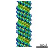 2937MC 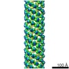 2936C 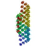 4uf8C C: citing same article ( M: map data used to model this data |
|---|---|
| Similar structure data |
-Links
 PDBj
PDBj


- Assembly
| Deposited unit | 
|
|---|---|
| 1 | x 20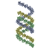
|
| 2 |
|
| 3 | 
|
| Symmetry | Helical symmetry: (Circular symmetry: 1 / Dyad axis: no / N subunits divisor: 1 / Num. of operations: 20 / Rise per n subunits: 10.09 Å / Rotation per n subunits: -26.71 °) |
-Components
| #1: Protein | Mass: 13704.609 Da / Num. of mol.: 3 / Fragment: PB1 DOMAIN, RESIDUES 1-122 Source method: isolated from a genetically manipulated source Source: (gene. exp.) HOMO SAPIENS (human) / Plasmid: POPTM-P62-PB1 / Production host:  |
|---|
-Experimental details
-Experiment
| Experiment | Method: ELECTRON MICROSCOPY |
|---|---|
| EM experiment | Aggregation state: FILAMENT / 3D reconstruction method: helical reconstruction |
- Sample preparation
| Component | Name: PB1(1-122) DOMAIN OF P62- SQSTM-1 / Type: COMPLEX |
|---|---|
| Buffer solution | Name: 50 MM TRIS PH 7.5, 100 MM NACL, DTT 4 MM / pH: 7.5 / Details: 50 MM TRIS PH 7.5, 100 MM NACL, DTT 4 MM |
| Specimen | Conc.: 0.25 mg/ml / Embedding applied: NO / Shadowing applied: NO / Staining applied: NO / Vitrification applied: YES |
| Specimen support | Details: HOLEY CARBON |
| Vitrification | Instrument: HOMEMADE PLUNGER / Cryogen name: ETHANE Details: VITRIFICATION 1 -- CRYOGEN- ETHANE, TEMPERATURE- 77, INSTRUMENT- HOMEMADE PLUNGER, METHOD- BACKSIDE BLOTTING, |
- Electron microscopy imaging
| Experimental equipment |  Model: Titan Krios / Image courtesy: FEI Company |
|---|---|
| Microscopy | Model: FEI TITAN KRIOS / Date: Oct 9, 2012 |
| Electron gun | Electron source:  FIELD EMISSION GUN / Accelerating voltage: 120 kV / Illumination mode: FLOOD BEAM FIELD EMISSION GUN / Accelerating voltage: 120 kV / Illumination mode: FLOOD BEAM |
| Electron lens | Mode: BRIGHT FIELD / Nominal magnification: 59000 X / Nominal defocus max: 3500 nm / Nominal defocus min: 1500 nm / Cs: 2.7 mm |
| Image recording | Electron dose: 10 e/Å2 / Film or detector model: GATAN ULTRASCAN 4000 (4k x 4k) |
| Image scans | Num. digital images: 443 |
| Radiation wavelength | Relative weight: 1 |
- Processing
| EM software |
| ||||||||||||
|---|---|---|---|---|---|---|---|---|---|---|---|---|---|
| CTF correction | Details: CTFFIND, CONVOLUTION IMAGES, WIENER FILTER RECONSTRUCTION | ||||||||||||
| 3D reconstruction | Method: PROJECTION MATCHING / Resolution: 10.3 Å / Num. of particles: 182238 / Nominal pixel size: 1.372 Å / Actual pixel size: 1.372 Å Details: SUBMISSION BASED ON EXPERIMENTAL DATA FROM EMDB EMD-2937. (DEPOSITION ID: 13204). Symmetry type: HELICAL | ||||||||||||
| Atomic model building | Protocol: RIGID BODY FIT / Space: REAL / Details: METHOD--RIGID BODY REFINEMENT PROTOCOL--NMR | ||||||||||||
| Atomic model building | PDB-ID: 2KKC Accession code: 2KKC / Source name: PDB / Type: experimental model | ||||||||||||
| Refinement | Highest resolution: 10.3 Å | ||||||||||||
| Refinement step | Cycle: LAST / Highest resolution: 10.3 Å
|

