 Movie
Movie Controller

[English] 日本語
 Yorodumi
- PDB-1tzn: Crystal Structure of the Anthrax Toxin Protective Antigen Heptame... -
+ Open data

- Basic information
| Entry | Database: PDB / ID: 1tzn | ||||||
|---|---|---|---|---|---|---|---|
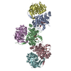
| Title | Crystal Structure of the Anthrax Toxin Protective Antigen Heptameric Prepore bound to the VWA domain of CMG2, an anthrax toxin receptor | ||||||
 Components Components |
| ||||||
 Keywords Keywords | TOXIN RECEPTOR/TOXIN / heptamer / TOXIN RECEPTOR-TOXIN COMPLEX | ||||||
| Function / homology |  Function and homology information Function and homology informationsymbiont-mediated suppression of host MAPK cascade / collagen fibril organization / host cell cytosol / uterus development / Uptake and function of anthrax toxins / single fertilization / host cell endosome membrane / protein homooligomerization / transmembrane signaling receptor activity / toxin activity ...symbiont-mediated suppression of host MAPK cascade / collagen fibril organization / host cell cytosol / uterus development / Uptake and function of anthrax toxins / single fertilization / host cell endosome membrane / protein homooligomerization / transmembrane signaling receptor activity / toxin activity / endosome membrane / external side of plasma membrane / endoplasmic reticulum membrane / host cell plasma membrane / cell surface / extracellular region / metal ion binding / identical protein binding / plasma membrane Similarity search - Function | ||||||
| Biological species |   Homo sapiens (human) Homo sapiens (human) | ||||||
| Method |  X-RAY DIFFRACTION / SYNCHROTRON / MOLECULAR REPLACEMENT / Resolution: 4.3 Å X-RAY DIFFRACTION / SYNCHROTRON / MOLECULAR REPLACEMENT / Resolution: 4.3 Å | ||||||
 Authors Authors | Lacy, D.B. / Wigelsworth, D.J. / Melnyk, R.A. / Collier, R.J. | ||||||
 Citation Citation | Journal: Proc.Natl.Acad.Sci.USA / Year: 2004 Title: Structure of heptameric protective antigen bound to an anthrax toxin receptor: A role for receptor in pH-dependent pore formation Authors: Lacy, D.B. / Wigelsworth, D.J. / Melnyk, R.A. / Harrison, S.C. / Collier, R.J. | ||||||
| History |
|
- Structure visualization
| Structure viewer | Molecule: MolmilJmol/JSmol |
|---|
- Downloads & links
-Download
| PDBx/mmCIF format | 1tzn.cif.gz | 1.8 MB | Display | PDBx/mmCIF format |
|---|---|---|---|---|
| PDB format | pdb1tzn.ent.gz | 1.5 MB | Display | PDB format |
| PDBx/mmJSON format | 1tzn.json.gz | Tree view | PDBx/mmJSON format | |
| Others |  Other downloads Other downloads |
-Validation report
| Arichive directory | https://data.pdbj.org/pub/pdb/validation_reports/tz/1tznftp://data.pdbj.org/pub/pdb/validation_reports/tz/1tzn | HTTPS FTP |
|---|
-Related structure data


-Links
 PDBj
PDBj


- Assembly
| Deposited unit | 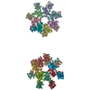
| ||||||||||
|---|---|---|---|---|---|---|---|---|---|---|---|
| 1 | 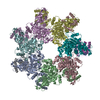
| ||||||||||
| 2 | 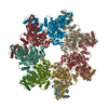
| ||||||||||
| 3 | 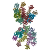
| ||||||||||
| 4 | 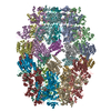
| ||||||||||
| 5 | 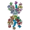
| ||||||||||
| Unit cell |
| ||||||||||
| Details | The biological assembly is one heptamer |
-Components
| #1: Protein | Mass: 63019.012 Da / Num. of mol.: 14 / Fragment: 63-kDa domain Source method: isolated from a genetically manipulated source Source: (gene. exp.) #2: Protein | Mass: 19840.791 Da / Num. of mol.: 14 / Fragment: VWA domain Source method: isolated from a genetically manipulated source Source: (gene. exp.) Homo sapiens (human) / Gene: ANTXR2 / Plasmid: pGEX-4T-1 / Production host: #3: Chemical | ChemComp-CA /   Mass: 40.078 Da / Num. of mol.: 28 / Source method: obtained synthetically / Formula: Ca Mass: 40.078 Da / Num. of mol.: 28 / Source method: obtained synthetically / Formula: Ca#4: Chemical | ChemComp-MG /   Mass: 24.305 Da / Num. of mol.: 14 / Source method: obtained synthetically / Formula: Mg Mass: 24.305 Da / Num. of mol.: 14 / Source method: obtained synthetically / Formula: MgHas protein modification | Y | |
|---|
-Experimental details
-Experiment
| Experiment | Method: X-RAY DIFFRACTION / Number of used crystals: 1 |
|---|
- Sample preparation
| Crystal | Density Matthews: 3.8 Å3/Da / Density % sol: 63 % |
|---|---|
| Crystal grow | Temperature: 277 K / Method: vapor diffusion, hanging drop / pH: 8.5 Details: PEG 8000, sodium chloride, Tris, magnesium chloride, pH 8.5, VAPOR DIFFUSION, HANGING DROP, temperature 277K |
-Data collection
| Diffraction | Mean temperature: 100 K |
|---|---|
| Diffraction source | Source: SYNCHROTRON / Site: CHESS  / Beamline: F1 / Wavelength: 0.9186 Å / Beamline: F1 / Wavelength: 0.9186 Å |
| Detector | Type: ADSC QUANTUM 4 / Detector: CCD / Date: Mar 15, 2004 / Details: null |
| Radiation | Monochromator: horizontally bent Si(III) / Protocol: SINGLE WAVELENGTH / Monochromatic (M) / Laue (L): M / Scattering type: x-ray |
| Radiation wavelength | Wavelength: 0.9186 Å / Relative weight: 1 |
| Reflection | Resolution: 4.3→20 Å / Num. all: 120016 / Num. obs: 120016 / % possible obs: 97.3 % / Observed criterion σ(I): 1.8 / Redundancy: 2.7 % / Rsym value: 0.165 / Net I/σ(I): 6.8 |
| Reflection shell | Resolution: 4.3→4.45 Å / Redundancy: 2.5 % / Mean I/σ(I) obs: 1.8 / Num. unique all: 12124 / Rsym value: 0.449 / % possible all: 98.1 |
- Processing
| Software |
| |||||||||||||||||||||||||
|---|---|---|---|---|---|---|---|---|---|---|---|---|---|---|---|---|---|---|---|---|---|---|---|---|---|---|
| Refinement | Method to determine structure: MOLECULAR REPLACEMENT Starting model: 3.6 heptameric prepore model Resolution: 4.3→20 Å / Cross valid method: THROUGHOUT / σ(F): 1 / Stereochemistry target values: Engh & Huber
| |||||||||||||||||||||||||
| Refinement step | Cycle: LAST / Resolution: 4.3→20 Å
| |||||||||||||||||||||||||
| Refine LS restraints |
|
