 Movie
Movie Controller


+ Open data

- Basic information
| Entry | Database: PDB / ID: 4uby | ||||||
|---|---|---|---|---|---|---|---|
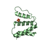
| Title | Crystal structure of a polymorphic beta1 peptide | ||||||
 Components Components | prion peptide | ||||||
 Keywords Keywords | de novo protein / membrane protein / prion peptide | ||||||
| Biological species | synthetic construct (others) | ||||||
| Method |  X-RAY DIFFRACTION / SYNCHROTRON / AB INITIO PHASING / Resolution: 1 Å X-RAY DIFFRACTION / SYNCHROTRON / AB INITIO PHASING / Resolution: 1 Å | ||||||
 Authors Authors | Yu, L. / Lee, S.-J. / Yee, V. | ||||||
 Citation Citation | Journal: Biochemistry / Year: 2015 Title: Crystal Structures of Polymorphic Prion Protein beta 1 Peptides Reveal Variable Steric Zipper Conformations. Authors: Yu, L. / Lee, S.J. / Yee, V.C. | ||||||
| History |
|
- Structure visualization
| Structure viewer | Molecule:  MolmilJmol/JSmol MolmilJmol/JSmol |
|---|
- Downloads & links
-Download
| PDBx/mmCIF format | 4uby.cif.gz | 17.8 KB | Display | PDBx/mmCIF format |
|---|---|---|---|---|
| PDB format | pdb4uby.ent.gz | 11.4 KB | Display | PDB format |
| PDBx/mmJSON format | 4uby.json.gz | Tree view | PDBx/mmJSON format | |
| Others |  Other downloads Other downloads |
-Validation report
| Arichive directory | https://data.pdbj.org/pub/pdb/validation_reports/ub/4ubyftp://data.pdbj.org/pub/pdb/validation_reports/ub/4uby | HTTPS FTP |
|---|
-Related structure data
| Related structure data |  4tutC  4ubzC  4w5lC  4w5mC 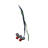 4w5pC 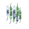 4w5yC 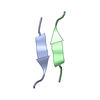 4w67C 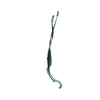 4w71C  4wbuC  4wbvC C: citing same article ( |
|---|---|
| Similar structure data |


-Links
 PDBj
PDBj
- Assembly
| Deposited unit | 
| ||||||||
|---|---|---|---|---|---|---|---|---|---|
| 1 | 
| ||||||||
| Unit cell |
| ||||||||
| Details | biological unit displays only a portion of the crystal lattice to demonstrate the crystal packing content. The crystal packing is formed by a repetition in both directions of the portion indicated in remark 350. |
-Components
| #1: Protein/peptide | Mass: 564.633 Da / Num. of mol.: 4 / Source method: obtained synthetically / Source: (synth.) synthetic construct (others) #2: Water | ChemComp-HOH / |  Mass: 18.015 Da / Num. of mol.: 22 / Source method: isolated from a natural source / Formula: H2O Mass: 18.015 Da / Num. of mol.: 22 / Source method: isolated from a natural source / Formula: H2O |
|---|
-Experimental details
-Experiment
| Experiment | Method: X-RAY DIFFRACTION / Number of used crystals: 1 |
|---|
- Sample preparation
| Crystal | Density Matthews: 1.66 Å3/Da / Density % sol: 25.86 % |
|---|---|
| Crystal grow | Temperature: 293 K / Method: vapor diffusion, sitting drop / pH: 4.6 / Details: 0.1 M sodium acetate, and 2 M ammonium sulfate |
-Data collection
| Diffraction | Mean temperature: 100 K | ||||||||||||||||||||||||||||||||||||||||||||||||||||||||||||||||||
|---|---|---|---|---|---|---|---|---|---|---|---|---|---|---|---|---|---|---|---|---|---|---|---|---|---|---|---|---|---|---|---|---|---|---|---|---|---|---|---|---|---|---|---|---|---|---|---|---|---|---|---|---|---|---|---|---|---|---|---|---|---|---|---|---|---|---|---|
| Diffraction source | Source: SYNCHROTRON / Site: APS  / Beamline: 23-ID-B / Wavelength: 0.7749 Å / Beamline: 23-ID-B / Wavelength: 0.7749 Å | ||||||||||||||||||||||||||||||||||||||||||||||||||||||||||||||||||
| Detector | Type: MARMOSAIC 300 mm CCD / Detector: CCD / Date: Oct 20, 2011 | ||||||||||||||||||||||||||||||||||||||||||||||||||||||||||||||||||
| Radiation | Protocol: SINGLE WAVELENGTH / Monochromatic (M) / Laue (L): M / Scattering type: x-ray | ||||||||||||||||||||||||||||||||||||||||||||||||||||||||||||||||||
| Radiation wavelength | Wavelength: 0.7749 Å / Relative weight: 1 | ||||||||||||||||||||||||||||||||||||||||||||||||||||||||||||||||||
| Reflection | Resolution: 1→50 Å / Num. obs: 6346 / % possible obs: 80.9 % / Redundancy: 2.8 % / Biso Wilson estimate: 2.67 Å2 / Rmerge(I) obs: 0.062 / Χ2: 1.016 / Net I/av σ(I): 16.26 / Net I/σ(I): 24.6 / Num. measured all: 18020 | ||||||||||||||||||||||||||||||||||||||||||||||||||||||||||||||||||
| Reflection shell | Diffraction-ID: 1 / Rejects: _
|
- Processing
| Software |
| ||||||||||||||||||||||||
|---|---|---|---|---|---|---|---|---|---|---|---|---|---|---|---|---|---|---|---|---|---|---|---|---|---|
| Refinement | Method to determine structure: AB INITIO PHASING / Resolution: 1→13.822 Å / SU ML: 0.06 / Cross valid method: FREE R-VALUE / σ(F): 2.32 / Phase error: 16.86 / Stereochemistry target values: ML
| ||||||||||||||||||||||||
| Solvent computation | Shrinkage radii: 0.9 Å / VDW probe radii: 1.11 Å / Solvent model: FLAT BULK SOLVENT MODEL | ||||||||||||||||||||||||
| Displacement parameters | Biso max: 34.08 Å2 / Biso mean: 5.8555 Å2 / Biso min: 1.64 Å2 | ||||||||||||||||||||||||
| Refinement step | Cycle: final / Resolution: 1→13.822 Å
| ||||||||||||||||||||||||
| Refine LS restraints |
| ||||||||||||||||||||||||
| LS refinement shell | Refine-ID: X-RAY DIFFRACTION / Total num. of bins used: 2
|
