 Movie
Movie Controller


+ Open data

- Basic information
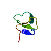
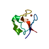
| Entry | Database: PDB / ID: 4wbu | ||||||
|---|---|---|---|---|---|---|---|
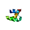
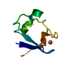
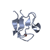
| Title | prion peptide | ||||||
 Components Components | PrP peptide | ||||||
 Keywords Keywords | de novo protein / membrane protein / prion peptide | ||||||
| Biological species | synthetic construct (others) | ||||||
| Method |  X-RAY DIFFRACTION / SYNCHROTRON / AB INITIO PHASING / Resolution: 1.15 Å X-RAY DIFFRACTION / SYNCHROTRON / AB INITIO PHASING / Resolution: 1.15 Å | ||||||
 Authors Authors | Yu, L. / Lee, S.-J. / Yee, V. | ||||||
 Citation Citation | Journal: Biochemistry / Year: 2015 Title: Crystal Structures of Polymorphic Prion Protein beta 1 Peptides Reveal Variable Steric Zipper Conformations. Authors: Yu, L. / Lee, S.J. / Yee, V.C. | ||||||
| History |
|
- Structure visualization
| Structure viewer | Molecule:  MolmilJmol/JSmol MolmilJmol/JSmol |
|---|
- Downloads & links
-Download
| PDBx/mmCIF format | 4wbu.cif.gz | 13.7 KB | Display | PDBx/mmCIF format |
|---|---|---|---|---|
| PDB format | pdb4wbu.ent.gz | 7.8 KB | Display | PDB format |
| PDBx/mmJSON format | 4wbu.json.gz | Tree view | PDBx/mmJSON format | |
| Others |  Other downloads Other downloads |
-Validation report
| Arichive directory | https://data.pdbj.org/pub/pdb/validation_reports/wb/4wbuftp://data.pdbj.org/pub/pdb/validation_reports/wb/4wbu | HTTPS FTP |
|---|
-Related structure data
| Related structure data | 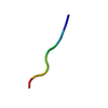 4tutC  4ubyC 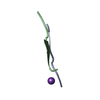 4ubzC 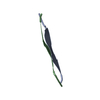 4w5lC 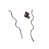 4w5mC  4w5pC  4w5yC  4w67C  4w71C  4wbvC C: citing same article ( |
|---|---|
| Similar structure data |


-Links
 PDBj
PDBj
- Assembly
| Deposited unit | 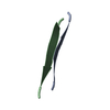
| ||||||||
|---|---|---|---|---|---|---|---|---|---|
| 1 | 
| ||||||||
| Unit cell |
| ||||||||
| Details | BIOLOGICAL UNIT DISPLAYS ONLY A PORTION OF THE CRYSTAL LATTICE TO DEMONSTRATE THE CRYSTAL PACKING CONTENT. THE CRYSTAL PACKING IS FORMED BY A REPETITION IN BOTH DIRECTIONS OF THE PORTION INDICATED IN REMARK 350. |
-Components
| #1: Protein/peptide | Mass: 626.724 Da / Num. of mol.: 2 / Source method: obtained synthetically / Details: synthetic / Source: (synth.) synthetic construct (others) #2: Water | ChemComp-HOH / |  Mass: 18.015 Da / Num. of mol.: 9 / Source method: isolated from a natural source / Formula: H2O Mass: 18.015 Da / Num. of mol.: 9 / Source method: isolated from a natural source / Formula: H2OHas protein modification | N | |
|---|
-Experimental details
-Experiment
| Experiment | Method: X-RAY DIFFRACTION / Number of used crystals: 1 |
|---|
- Sample preparation
| Crystal | Density Matthews: 1.49 Å3/Da / Density % sol: 17.59 % |
|---|---|
| Crystal grow | Temperature: 293 K / Method: vapor diffusion, sitting drop / pH: 7.5 Details: 0.1 M Hepes, 2.0 M ammonium sulfate, and 2.0 M NDSB-211 |
-Data collection
| Diffraction | Mean temperature: 100 K | ||||||||||||||||||||||||||||||||||||||||||||||||||||||||||||||||||
|---|---|---|---|---|---|---|---|---|---|---|---|---|---|---|---|---|---|---|---|---|---|---|---|---|---|---|---|---|---|---|---|---|---|---|---|---|---|---|---|---|---|---|---|---|---|---|---|---|---|---|---|---|---|---|---|---|---|---|---|---|---|---|---|---|---|---|---|
| Diffraction source | Source: SYNCHROTRON / Site: APS  / Beamline: 19-ID / Wavelength: 0.9184 Å / Beamline: 19-ID / Wavelength: 0.9184 Å | ||||||||||||||||||||||||||||||||||||||||||||||||||||||||||||||||||
| Detector | Type: ADSC QUANTUM 315 / Detector: CCD / Date: Mar 31, 2007 | ||||||||||||||||||||||||||||||||||||||||||||||||||||||||||||||||||
| Radiation | Protocol: SINGLE WAVELENGTH / Monochromatic (M) / Laue (L): M / Scattering type: x-ray | ||||||||||||||||||||||||||||||||||||||||||||||||||||||||||||||||||
| Radiation wavelength | Wavelength: 0.9184 Å / Relative weight: 1 | ||||||||||||||||||||||||||||||||||||||||||||||||||||||||||||||||||
| Reflection | Resolution: 1.15→50 Å / Num. obs: 2627 / % possible obs: 87.6 % / Redundancy: 5.6 % / Biso Wilson estimate: 3.56 Å2 / Rmerge(I) obs: 0.102 / Χ2: 1.001 / Net I/av σ(I): 11.957 / Net I/σ(I): 11.1 / Num. measured all: 14648 | ||||||||||||||||||||||||||||||||||||||||||||||||||||||||||||||||||
| Reflection shell | Diffraction-ID: 1 / Rejects: _
|
- Processing
| Software |
| ||||||||||||||||||||||||
|---|---|---|---|---|---|---|---|---|---|---|---|---|---|---|---|---|---|---|---|---|---|---|---|---|---|
| Refinement | Method to determine structure: AB INITIO PHASING / Resolution: 1.15→22.28 Å / SU ML: 0.04 / Cross valid method: FREE R-VALUE / σ(F): 1.46 / Phase error: 16.74 / Stereochemistry target values: ML
| ||||||||||||||||||||||||
| Solvent computation | Shrinkage radii: 0.9 Å / VDW probe radii: 1.11 Å / Solvent model: FLAT BULK SOLVENT MODEL | ||||||||||||||||||||||||
| Displacement parameters | Biso max: 35.49 Å2 / Biso mean: 8.2041 Å2 / Biso min: 2.25 Å2 | ||||||||||||||||||||||||
| Refinement step | Cycle: final / Resolution: 1.15→22.28 Å
| ||||||||||||||||||||||||
| Refine LS restraints |
| ||||||||||||||||||||||||
| LS refinement shell | Resolution: 1.1504→22.2847 Å / Total num. of bins used: 1
|
