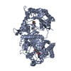
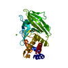
Entry Database : PDB / ID : 4u10Title Probing the structure and mechanism of de-N-acetylase from aggregatibacter actinomycetemcomitans Poly-beta-1,6-N-acetyl-D-glucosamine N-deacetylase Keywords / / / Function / homology Function Domain/homology Component
/ / / / / / / / / / / / / / / / / / / / / / / Biological species Aggregatibacter actinomycetemcomitans (bacteria)Method / / Resolution : 2.05 Å Authors Varudharasu, D. / Narayanan, R. Funding support Organization Grant number Country United States Public Health Service DE22544
Journal : To Be Published Title : Probing the structure and mechanism of de-N-acetylase from aggregatibacter actinomycetemcomitansAuthors : Varudharsu, D. / Narayanan, R. History Deposition Jul 14, 2014 Deposition site / Processing site Revision 1.0 Jan 14, 2015 Provider / Type Revision 1.1 Sep 27, 2023 Group Data collection / Database references ... Data collection / Database references / Derived calculations / Other / Refinement description / Source and taxonomy / Structure summary Category chem_comp_atom / chem_comp_bond ... chem_comp_atom / chem_comp_bond / citation / database_2 / entity_src_gen / pdbx_database_status / pdbx_initial_refinement_model / pdbx_struct_assembly / pdbx_struct_conn_angle / pdbx_struct_oper_list / refine_hist / struct_conn / struct_keywords Item _citation.journal_id_CSD / _database_2.pdbx_DOI ... _citation.journal_id_CSD / _database_2.pdbx_DOI / _database_2.pdbx_database_accession / _entity_src_gen.pdbx_alt_source_flag / _pdbx_database_status.pdb_format_compatible / _pdbx_struct_assembly.oligomeric_details / _pdbx_struct_conn_angle.ptnr1_auth_asym_id / _pdbx_struct_conn_angle.ptnr1_auth_comp_id / _pdbx_struct_conn_angle.ptnr1_auth_seq_id / _pdbx_struct_conn_angle.ptnr1_label_asym_id / _pdbx_struct_conn_angle.ptnr1_label_atom_id / _pdbx_struct_conn_angle.ptnr1_label_comp_id / _pdbx_struct_conn_angle.ptnr1_label_seq_id / _pdbx_struct_conn_angle.ptnr2_auth_seq_id / _pdbx_struct_conn_angle.ptnr2_label_alt_id / _pdbx_struct_conn_angle.ptnr2_label_asym_id / _pdbx_struct_conn_angle.ptnr2_symmetry / _pdbx_struct_conn_angle.ptnr3_auth_asym_id / _pdbx_struct_conn_angle.ptnr3_auth_comp_id / _pdbx_struct_conn_angle.ptnr3_auth_seq_id / _pdbx_struct_conn_angle.ptnr3_label_asym_id / _pdbx_struct_conn_angle.ptnr3_label_atom_id / _pdbx_struct_conn_angle.ptnr3_label_comp_id / _pdbx_struct_conn_angle.ptnr3_label_seq_id / _pdbx_struct_conn_angle.value / _pdbx_struct_oper_list.symmetry_operation / _refine_hist.number_atoms_solvent / _refine_hist.number_atoms_total / _refine_hist.pdbx_number_atoms_ligand / _refine_hist.pdbx_number_atoms_nucleic_acid / _refine_hist.pdbx_number_atoms_protein / _struct_conn.conn_type_id / _struct_conn.id / _struct_conn.pdbx_dist_value / _struct_conn.pdbx_leaving_atom_flag / _struct_conn.pdbx_ptnr2_label_alt_id / _struct_conn.ptnr1_auth_asym_id / _struct_conn.ptnr1_auth_comp_id / _struct_conn.ptnr1_auth_seq_id / _struct_conn.ptnr1_label_asym_id / _struct_conn.ptnr1_label_atom_id / _struct_conn.ptnr1_label_comp_id / _struct_conn.ptnr1_label_seq_id / _struct_conn.ptnr2_auth_asym_id / _struct_conn.ptnr2_auth_comp_id / _struct_conn.ptnr2_auth_seq_id / _struct_conn.ptnr2_label_asym_id / _struct_conn.ptnr2_label_atom_id / _struct_conn.ptnr2_label_comp_id / _struct_conn.ptnr2_label_seq_id / _struct_conn.ptnr2_symmetry / _struct_keywords.text Revision 1.2 Nov 15, 2023 Group / Category / chem_comp_bond / Item / _chem_comp_bond.atom_id_2Revision 1.3 Oct 16, 2024 Group / Category / pdbx_modification_feature
Show all Show less
 Movie
Movie Controller

 Yorodumi
 Open data
 Basic information
 Components
 Keywords
 Function and homology information
 Aggregatibacter actinomycetemcomitans (bacteria)
 X-RAY DIFFRACTION /
  Authors
 United States, 1items
  Citation
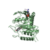
 Structure visualization
 Downloads & links
 Other downloads
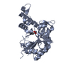


 PDBj
PDBj
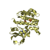
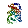
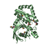
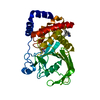
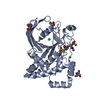
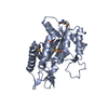
 Assembly
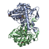


 Mass: 65.409 Da / Num. of mol.: 7 / Source method: obtained synthetically / Formula: Zn
 Mass: 35.453 Da / Num. of mol.: 2 / Source method: obtained synthetically / Formula: Cl
 Mass: 18.015 Da / Num. of mol.: 258 / Source method: isolated from a natural source / Formula: H2O
 Sample preparation
 Processing