 Movie
Movie Controller

[English] 日本語
 Yorodumi
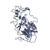
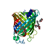
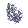
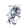
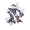
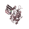
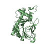
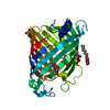
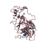
- PDB-3mo2: human G9a-like (GLP, also known as EHMT1) in complex with inhibit... -
+ Open data

- Basic information
| Entry | Database: PDB / ID: 3mo2 | ||||||
|---|---|---|---|---|---|---|---|
| Title | human G9a-like (GLP, also known as EHMT1) in complex with inhibitor E67 | ||||||
 Components Components | Histone-lysine N-methyltransferase, H3 lysine-9 specific 5 | ||||||
 Keywords Keywords | TRANSFERASE / Epigenetics / Histone lysine methylation / enzymatic inhibition / lysine mimics | ||||||
| Function / homology |  Function and homology information Function and homology informationnegative regulation of white fat cell differentiation / histone H3K9me2 methyltransferase activity / histone H3K9 trimethyltransferase activity / [histone H3]-lysine9 N-methyltransferase / peptidyl-lysine monomethylation / peptidyl-lysine dimethylation / histone H3K9 methyltransferase activity / histone H3K9 monomethyltransferase activity / histone H3K27 methyltransferase activity / facultative heterochromatin formation ...negative regulation of white fat cell differentiation / histone H3K9me2 methyltransferase activity / histone H3K9 trimethyltransferase activity / [histone H3]-lysine9 N-methyltransferase / peptidyl-lysine monomethylation / peptidyl-lysine dimethylation / histone H3K9 methyltransferase activity / histone H3K9 monomethyltransferase activity / histone H3K27 methyltransferase activity / facultative heterochromatin formation / C2H2 zinc finger domain binding / protein-lysine N-methyltransferase activity / beige fat cell differentiation / DNA methylation-dependent constitutive heterochromatin formation / Transcriptional Regulation by E2F6 / Transcriptional Regulation by VENTX / regulation of embryonic development / response to fungicide / epigenetic regulation of gene expression / brown fat cell differentiation / Transferases; Transferring one-carbon groups; Methyltransferases / Regulation of endogenous retroelements by the Human Silencing Hub (HUSH) complex / transcription corepressor binding / methyltransferase activity / PKMTs methylate histone lysines / Regulation of TP53 Activity through Methylation / p53 binding / positive regulation of cold-induced thermogenesis / chromatin organization / Senescence-Associated Secretory Phenotype (SASP) / nuclear body / negative regulation of DNA-templated transcription / chromatin / negative regulation of transcription by RNA polymerase II / zinc ion binding / nucleoplasm / nucleus Similarity search - Function | ||||||
| Biological species |  Homo sapiens (human) Homo sapiens (human) | ||||||
| Method |  X-RAY DIFFRACTION / SYNCHROTRON / MOLECULAR REPLACEMENT / Resolution: 2.49 Å X-RAY DIFFRACTION / SYNCHROTRON / MOLECULAR REPLACEMENT / Resolution: 2.49 Å | ||||||
 Authors Authors | Chang, Y. / Horton, J.R. / Cheng, X. | ||||||
 Citation Citation | Journal: J.Mol.Biol. / Year: 2010 Title: Adding a lysine mimic in the design of potent inhibitors of histone lysine methyltransferases. Authors: Chang, Y. / Ganesh, T. / Horton, J.R. / Spannhoff, A. / Liu, J. / Sun, A. / Zhang, X. / Bedford, M.T. / Shinkai, Y. / Snyder, J.P. / Cheng, X. | ||||||
| History |
|
- Structure visualization
| Structure viewer | Molecule: MolmilJmol/JSmol |
|---|
- Downloads & links
-Download
| PDBx/mmCIF format | 3mo2.cif.gz | 232.7 KB | Display | PDBx/mmCIF format |
|---|---|---|---|---|
| PDB format | pdb3mo2.ent.gz | 182.3 KB | Display | PDB format |
| PDBx/mmJSON format | 3mo2.json.gz | Tree view | PDBx/mmJSON format | |
| Others |  Other downloads Other downloads |
-Validation report
| Arichive directory | https://data.pdbj.org/pub/pdb/validation_reports/mo/3mo2ftp://data.pdbj.org/pub/pdb/validation_reports/mo/3mo2 | HTTPS FTP |
|---|
-Related structure data
| Related structure data |  3mo0C  3mo5C  3fpdS S: Starting model for refinement C: citing same article ( |
|---|---|
| Similar structure data |

-Links
 PDBj
PDBj


- Assembly
| Deposited unit | 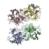
| ||||||||
|---|---|---|---|---|---|---|---|---|---|
| 1 | 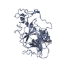
| ||||||||
| 2 | 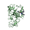
| ||||||||
| 3 | 
| ||||||||
| 4 | 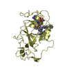
| ||||||||
| 5 | 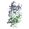
| ||||||||
| 6 | 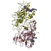
| ||||||||
| Unit cell |
| ||||||||
| Details | 4 complexes per asymmetric unit |
-Components
| #1: Protein | Mass: 32830.219 Da / Num. of mol.: 4 / Fragment: C-terminal SET domain Source method: isolated from a genetically manipulated source Source: (gene. exp.) Homo sapiens (human) / Gene: EHMT1, EUHMTASE1, KIAA1876, KMT1D / Production host:  References: UniProt: Q9H9B1, histone-lysine N-methyltransferase #2: Chemical | ChemComp-ZN /   Mass: 65.409 Da / Num. of mol.: 16 / Source method: obtained synthetically / Formula: Zn Mass: 65.409 Da / Num. of mol.: 16 / Source method: obtained synthetically / Formula: Zn#3: Chemical | ChemComp-E67 /   Mass: 549.751 Da / Num. of mol.: 4 / Source method: obtained synthetically / Formula: C31H47N7O2 Mass: 549.751 Da / Num. of mol.: 4 / Source method: obtained synthetically / Formula: C31H47N7O2#4: Chemical |   Type: L-peptide linking / Mass: 384.411 Da / Num. of mol.: 2 / Source method: obtained synthetically / Formula: C14H20N6O5S Type: L-peptide linking / Mass: 384.411 Da / Num. of mol.: 2 / Source method: obtained synthetically / Formula: C14H20N6O5S#5: Water | ChemComp-HOH / |  Mass: 18.015 Da / Num. of mol.: 397 / Source method: isolated from a natural source / Formula: H2O Mass: 18.015 Da / Num. of mol.: 397 / Source method: isolated from a natural source / Formula: H2O |
|---|
-Experimental details
-Experiment
| Experiment | Method: X-RAY DIFFRACTION / Number of used crystals: 1 |
|---|
- Sample preparation
| Crystal | Density Matthews: 2.54 Å3/Da / Density % sol: 51.51 % |
|---|---|
| Crystal grow | Temperature: 289 K / Method: vapor diffusion, hanging drop / pH: 7.5 Details: 0.1 M Hepes pH 7.5, 14% polyethylene glycol 4000, 9% isopropanol, and 12% dimethyl sulfoxide, VAPOR DIFFUSION, HANGING DROP, temperature 289K |
-Data collection
| Diffraction | Mean temperature: 100 K |
|---|---|
| Diffraction source | Source: SYNCHROTRON / Site: APS  / Beamline: 22-ID / Wavelength: 1 / Beamline: 22-ID / Wavelength: 1 |
| Detector | Type: MARMOSAIC 300 mm CCD / Detector: CCD / Date: Oct 14, 2009 |
| Radiation | Protocol: SINGLE WAVELENGTH / Monochromatic (M) / Laue (L): M / Scattering type: x-ray |
| Radiation wavelength | Wavelength: 1 Å / Relative weight: 1 |
| Reflection | Resolution: 2.49→31.83 Å / Num. obs: 43245 / % possible obs: 94.4 % / Observed criterion σ(F): 0 / Observed criterion σ(I): -3 / Redundancy: 4.3 % / Biso Wilson estimate: 27.9 Å2 / Rmerge(I) obs: 0.095 / Net I/σ(I): 12.8 |
| Reflection shell | Resolution: 2.49→2.58 Å / Redundancy: 4.2 % / Rmerge(I) obs: 0.384 / Mean I/σ(I) obs: 2.6 / % possible all: 94.3 |
- Processing
| Software |
| ||||||||||||||||||||||||||||||||||||||||||||||||||||||||||||||||||||||||||||||||
|---|---|---|---|---|---|---|---|---|---|---|---|---|---|---|---|---|---|---|---|---|---|---|---|---|---|---|---|---|---|---|---|---|---|---|---|---|---|---|---|---|---|---|---|---|---|---|---|---|---|---|---|---|---|---|---|---|---|---|---|---|---|---|---|---|---|---|---|---|---|---|---|---|---|---|---|---|---|---|---|---|---|
| Refinement | Method to determine structure: MOLECULAR REPLACEMENT Starting model: 3FPD Resolution: 2.49→31.83 Å / Rfactor Rfree error: 0.005 / Data cutoff high absF: 451338.01 / Data cutoff low absF: 0 / Isotropic thermal model: RESTRAINED / Cross valid method: THROUGHOUT / σ(F): 0 / Details: BULK SOLVENT MODEL USED
| ||||||||||||||||||||||||||||||||||||||||||||||||||||||||||||||||||||||||||||||||
| Solvent computation | Solvent model: FLAT MODEL / Bsol: 39.8381 Å2 / ksol: 0.35 e/Å3 | ||||||||||||||||||||||||||||||||||||||||||||||||||||||||||||||||||||||||||||||||
| Displacement parameters | Biso mean: 38.6 Å2
| ||||||||||||||||||||||||||||||||||||||||||||||||||||||||||||||||||||||||||||||||
| Refine analyze |
| ||||||||||||||||||||||||||||||||||||||||||||||||||||||||||||||||||||||||||||||||
| Refinement step | Cycle: LAST / Resolution: 2.49→31.83 Å
| ||||||||||||||||||||||||||||||||||||||||||||||||||||||||||||||||||||||||||||||||
| Refine LS restraints |
| ||||||||||||||||||||||||||||||||||||||||||||||||||||||||||||||||||||||||||||||||
| Refine LS restraints NCS | NCS model details: NONE | ||||||||||||||||||||||||||||||||||||||||||||||||||||||||||||||||||||||||||||||||
| LS refinement shell | Resolution: 2.49→2.58 Å / Rfactor Rfree error: 0.022 / Total num. of bins used: 10
| ||||||||||||||||||||||||||||||||||||||||||||||||||||||||||||||||||||||||||||||||
| Xplor file |
|
